Learn how magnetic resonance imaging (MRI) works, when it’s used in respiratory care, and key safety concerns for ventilated patients.
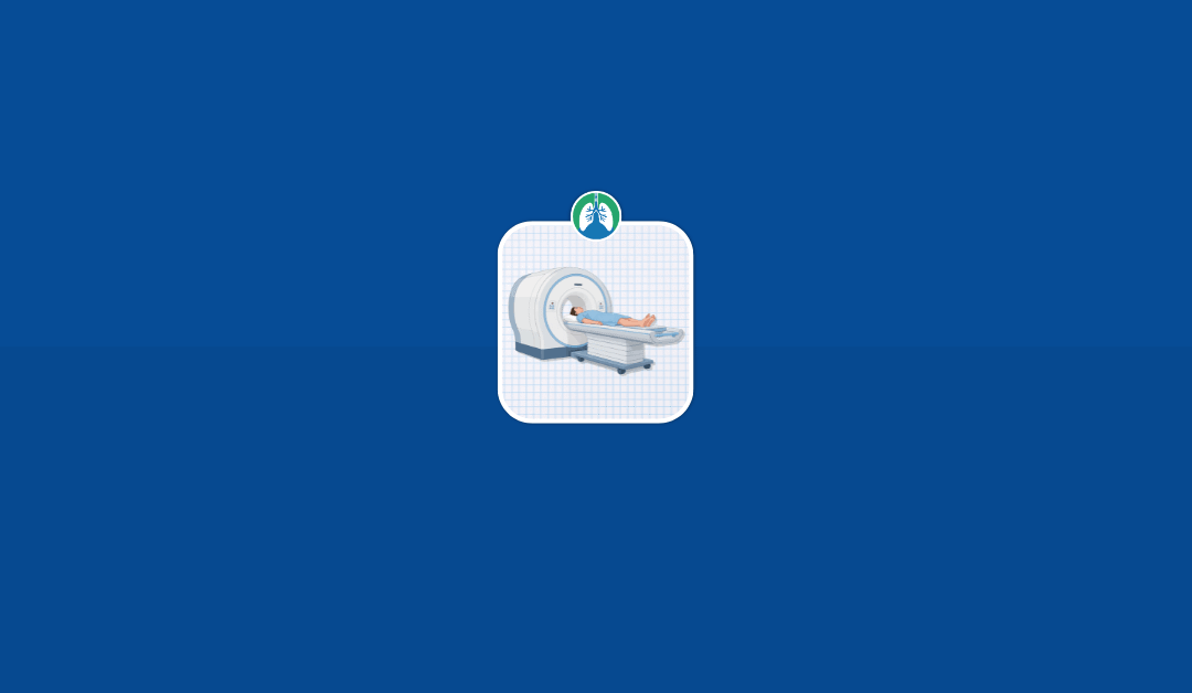

Learn how magnetic resonance imaging (MRI) works, when it’s used in respiratory care, and key safety concerns for ventilated patients.
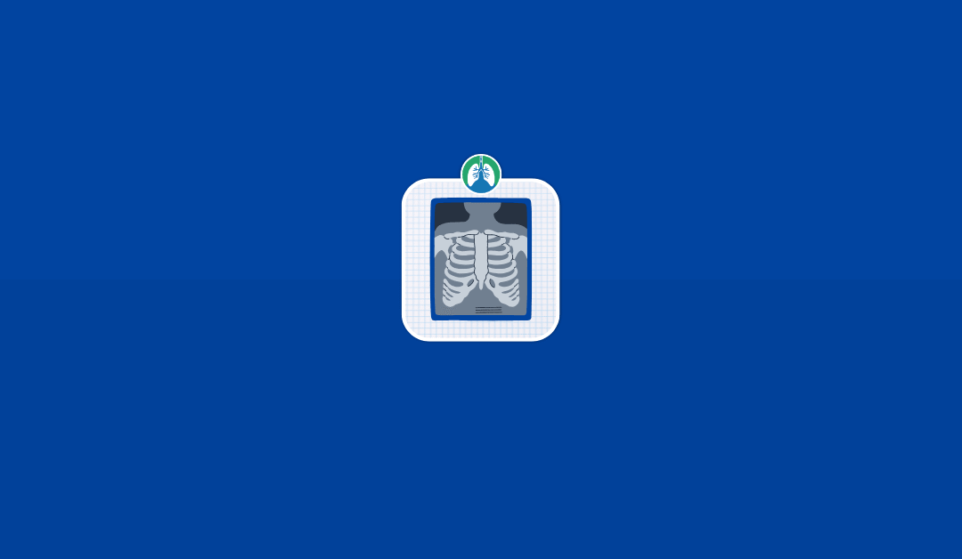
Learn radiographic findings, chest x-ray patterns, and key imaging clues used to assess lung conditions and guide respiratory care.
Learn how lateral neck radiographs help identify croup, epiglottitis, foreign bodies, enlarged adenoids, and upper-airway obstruction.
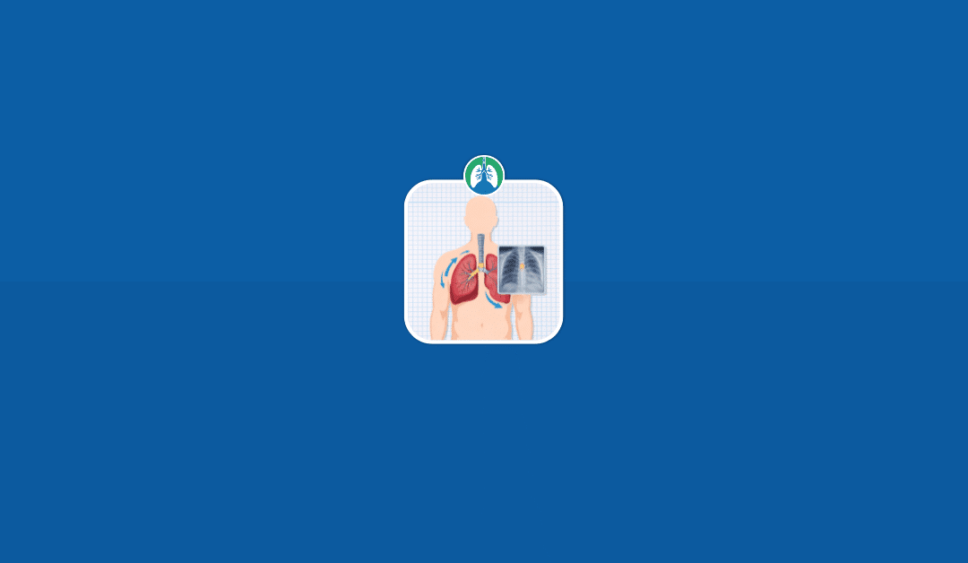
Learn how to identify foreign body aspiration on a chest X-ray, recognize key signs, and choose the right imaging for accurate diagnosis.
Learn what a ventilation-perfusion (V/Q) scan shows, how it helps detect a pulmonary embolism, and how the results are interpreted.
Learn how ultrasound is used in respiratory care, critical care, lung assessment, cardiac imaging, and bedside procedures.

Learn how computed tomography works and why it’s essential in respiratory care for diagnosing lung, pleural, and vascular diseases.
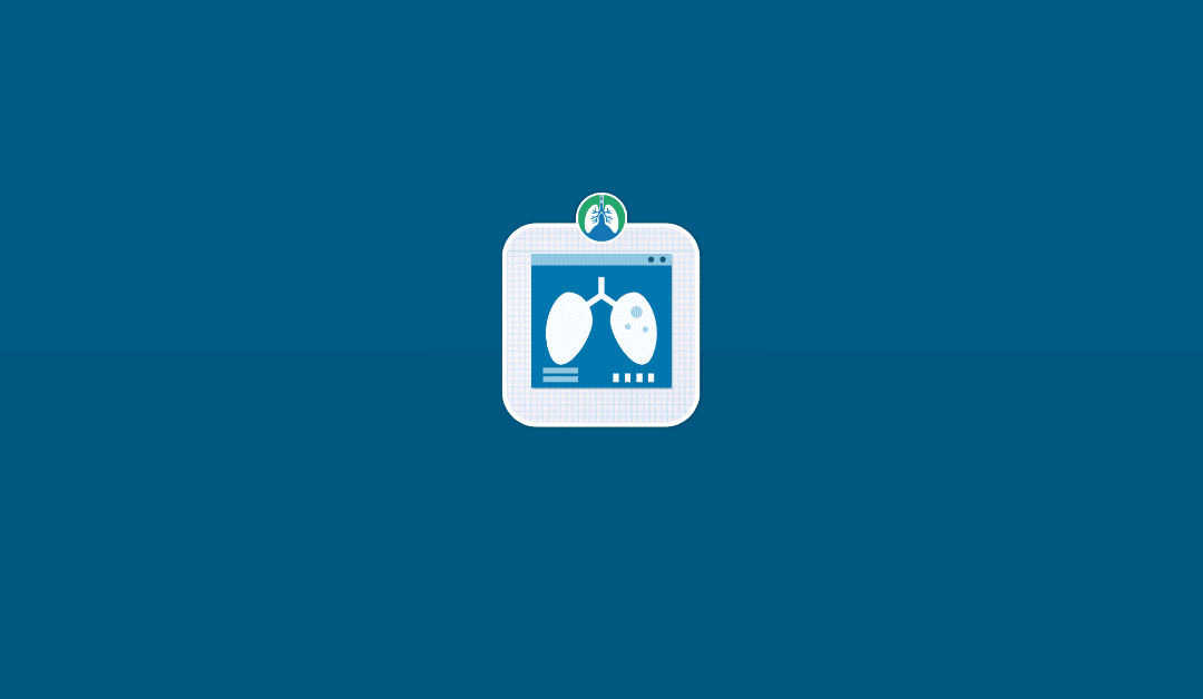
Learn what a chest radiograph (x-ray) is and why it’s essential for respiratory therapists in diagnosing and managing lung conditions.

Explore the types of thoracic imaging, their uses, and benefits for diagnosing chest conditions effectively in this comprehensive guide.

Learn about Kerley B lines, their causes, clinical significance, and what their presence on chest X-rays indicates for patient health.