
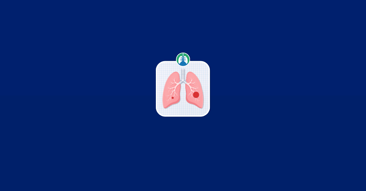
Finding out that an imaging test showed a spot on your lung can be unsettling, especially when the report uses terms such as “lung nodule” or “lung mass.” Although these words may sound similar, they have specific meanings that are largely based on size.
Neither term automatically means cancer. Lung nodules and masses can develop for many reasons, including infections, inflammation, scar tissue, and tumors.
Understanding the difference can make a radiology report easier to interpret and help you know what questions to ask your healthcare provider.

Download our free guide that has over 100+ of the best tips for healthy lungs.
What Is a Lung Nodule?
A lung nodule is a relatively small, rounded area of abnormal tissue seen within the lung. Radiologists generally define a pulmonary nodule as an opacity measuring less than 3 centimeters, or about 1.2 inches, in diameter.
Some nodules are only a few millimeters across and would never be noticed without modern imaging. They are commonly discovered on:
- Chest X-rays
- Computed tomography (CT) scans
- Low-dose CT scans performed for lung cancer screening
- Imaging performed for an unrelated medical problem
For example, someone may have a CT scan after a car accident, during evaluation for chest pain, or because of an infection. A small lung nodule may appear unexpectedly even though it has nothing to do with the original reason for the scan.
Many pulmonary nodules do not cause symptoms. In fact, their small size is one reason they are so frequently discovered incidentally.
A nodule describes what is seen on imaging. It does not describe what caused it. A pulmonary nodule may represent an old infection, a small area of inflammation, scar tissue, or a tumor, among other possibilities.
What Is a Lung Mass?
A lung mass is an abnormal area in the lung that is larger than a pulmonary nodule. In general, an abnormal lung lesion greater than 3 centimeters in diameter is referred to as a mass rather than a nodule.
The 3-centimeter distinction is primarily a radiologic definition. It does not mean that something measuring 2.9 centimeters is harmless while something measuring 3.1 centimeters is dangerous. Instead, size is one of several characteristics doctors use when estimating how likely an abnormality is to represent cancer or another condition.
A lung mass requires careful evaluation because larger lesions tend to carry greater concern for malignancy than very small nodules. However, a mass is still not a diagnosis of cancer.
Large areas of infection, inflammation, scar tissue, and other noncancerous processes can sometimes appear mass-like on imaging. Further testing is often needed before doctors can determine exactly what the abnormality represents.

Lung Mass vs. Nodule: The Main Difference
The simplest difference between a pulmonary nodule and a pulmonary mass is size. A lung nodule is generally less than 3 centimeters in diameter, while an abnormality larger than 3 centimeters is typically described as a lung mass.
This distinction is useful, but size alone does not tell doctors whether something is benign or malignant.
Doctors also consider:
- The patient’s age
- Smoking history
- Previous cancer history
- Size of the abnormality
- Whether it has grown
- Shape and borders
- Location within the lung
- Whether calcium is present
- Whether the lesion is solid or partly solid
- Findings elsewhere in the lungs or body
For this reason, two people with lung abnormalities of exactly the same size may receive very different recommendations.
A smooth, stable nodule in a younger person with a low risk of lung cancer may require nothing more than follow-up imaging. A similarly sized nodule with irregular borders that has grown in an older person with a significant smoking history could require a much more aggressive evaluation.
Does a Lung Nodule Mean Cancer?
No. Finding a pulmonary nodule does not mean that someone has lung cancer.
There are many possible causes of lung nodules, and benign causes are common. The American Lung Association notes that lung nodules can develop for several reasons and that doctors assess their appearance, growth, and the person’s overall cancer risk before deciding whether additional testing is necessary.
The likelihood that a particular nodule is cancerous depends heavily on its characteristics and the person in whom it is found. Very small nodules are generally less concerning than larger nodules, particularly when they have a benign appearance and remain unchanged over time.
Note: Doctors rarely make a diagnosis based solely on the fact that a nodule exists. Instead, they look at the entire picture.
Benign Causes of Lung Nodules
Several noncancerous conditions can create nodules. One common cause is a previous infection. When the immune system responds to certain infections, it may create a small area of inflammation called a granuloma.
Granulomas can sometimes remain visible long after the infection has resolved.
Examples of infectious causes may include:
- Histoplasmosis
- Tuberculosis
- Other fungal infections
- Certain bacterial infections
Exposure to some fungal organisms is more common in particular geographic regions, meaning a person’s location and travel history can sometimes help doctors interpret a finding. Other benign possibilities include scar tissue and inflammatory conditions.
A nodule may also represent a benign tumor, such as a hamartoma. Hamartomas are noncancerous growths composed of tissues normally found in the lung, arranged in an unusual way.
Certain calcification patterns or the presence of fat within a nodule may make a benign diagnosis more likely, although the final interpretation should always be made by a qualified healthcare professional.
Does a Lung Mass Mean Cancer?
A lung mass generally raises more concern than a small lung nodule because larger pulmonary abnormalities have a higher likelihood of representing a significant disease process. However, a lung mass does not automatically mean lung cancer.
Imaging terminology is descriptive. When a radiologist says there is a mass, that description is primarily telling the healthcare team that an abnormal area of a certain size and appearance is present. Doctors then have to determine its cause.
A lung mass could potentially result from:
- Lung cancer
- Cancer that has spread to the lung from another organ
- Pneumonia or another infection
- A lung abscess
- Fungal disease
- Tuberculosis
- Inflammatory disease
- Benign tumors
- Areas of organized inflammation or scar tissue
Note: Because cancer becomes an important possibility when a lung mass is identified, further evaluation is usually needed rather than assuming that the finding is harmless.
Why Size Matters
Size is an important part of evaluating any pulmonary lesion because the probability of cancer generally increases as a lesion becomes larger. That does not mean every large lesion is malignant or that every small lesion is benign.
Some lung cancers are discovered when they are only a few millimeters in diameter. At the same time, some relatively large abnormalities ultimately prove to be infections or benign growths.
The important point is that size changes the level of concern and often changes what doctors recommend next. For a very small nodule in a person with a low risk of lung cancer, observation with a future CT scan may be appropriate.
As a nodule becomes larger or develops suspicious features, doctors may consider additional testing, including PET/CT or biopsy. For certain incidentally detected nodules 6 millimeters or larger, follow-up CT and, depending on the clinical circumstances, PET/CT or tissue sampling may be appropriate.
Note: A pulmonary mass is already beyond the traditional 3-centimeter nodule threshold, so doctors generally investigate it more closely.
What Does a Suspicious Lung Nodule Look Like?
Radiologists examine much more than diameter when evaluating a pulmonary nodule. Certain characteristics may increase or decrease concern, although no single imaging feature provides a definite answer in every case.
Borders and Shape
A smooth, well-defined nodule may be less suspicious than one with irregular or spiculated edges. The word spiculated refers to thin strands or projections extending outward from the edge of a lesion.
Spiculation can occur with cancer, but imaging findings must always be interpreted in context.
Growth Over Time
One of the most valuable pieces of information is whether the nodule has changed. Doctors may compare a new CT scan with older scans from months or years earlier.
If an abnormality has remained unchanged for an appropriate period, that stability may make certain cancers less likely. Growth can increase concern, although infection and inflammation can also change in size.
This is why obtaining previous imaging can be extremely helpful. If you have had chest X-rays or CT scans at another hospital or clinic, your healthcare provider may ask for those images to be transferred for comparison.
Calcification
Some patterns of calcium within a nodule strongly favor a benign cause. Calcified granulomas, for example, can develop after previous infections. Not all calcification is automatically benign, however, so the actual pattern matters.
Density
Radiologists may describe a pulmonary nodule as:
- Solid
- Part-solid
- Ground-glass
Note: A ground-glass nodule appears hazy on CT while still allowing some underlying lung structures to remain visible. Different types of nodules have different possible causes and may require different follow-up schedules.
Location
The location of a nodule may also contribute to risk assessment. Some lung cancers occur more frequently in particular areas of the lungs, while certain infections or inflammatory conditions may follow different patterns.
Note: Location alone cannot determine the diagnosis.
Why Previous CT Scans Are So Important
When a new lung nodule or mass is discovered, one of the first things doctors may want to know is whether it is actually new. Previous scans can sometimes answer that question immediately.
Imagine that a CT scan today shows an 8-millimeter nodule. Without older imaging, doctors may need to monitor that nodule to see whether it changes.
If a CT performed four years earlier shows the same nodule at approximately the same size and with the same appearance, that information can significantly affect the assessment. This is why patients should tell their healthcare team about previous imaging, even if it was performed for an unrelated reason.
Old scans may exist from:
- Emergency department visits
- Pneumonia evaluations
- Heart testing
- Cancer screening
- Previous hospitalizations
- Trauma evaluations
- Preoperative testing
Note: Actual images are often more useful than simply reading the previous radiology report because a radiologist can directly compare the lesions.
Symptoms of a Lung Nodule or Mass
Small pulmonary nodules often cause no symptoms at all. Their discovery may come as a complete surprise. Larger lesions are also sometimes asymptomatic, but depending on their size, location, and underlying cause, symptoms can occur.
Possible symptoms associated with lung disease include:
- Persistent cough
- Coughing up blood
- Shortness of breath
- Chest discomfort
- Recurrent respiratory infections
- Wheezing
- Unexplained weight loss
- Loss of appetite
- Persistent fatigue
These symptoms are not specific to a lung mass or lung cancer. Many respiratory and nonrespiratory conditions can produce them.
A persistent cough, chest pain, coughing up blood, fever, or shortness of breath can be reasons for a healthcare provider to order chest imaging in the first place. Coughing up blood, significant breathing difficulty, or severe chest symptoms should receive prompt medical attention.
How Doctors Evaluate a Lung Nodule
There is no single test that is appropriate for every pulmonary nodule. The evaluation depends on the nodule itself and the person’s risk factors.
Reviewing the CT Scan
CT imaging provides much more detail than a standard chest X-ray and is particularly useful for evaluating small pulmonary abnormalities.
The radiologist may document:
- Exact size
- Location
- Shape
- Margins
- Density
- Calcification
- Number of nodules
- Changes compared with previous scans
Note: If the abnormality was initially noticed on a chest X-ray, a CT scan may be performed to characterize it more clearly.
Assessing Personal Risk
Doctors combine imaging information with the patient’s medical history.
Important factors may include:
- Age
- Current or previous tobacco use
- Amount and duration of smoking
- Family history of lung cancer
- Personal history of cancer
- Occupational exposures
- Previous infections
- Immune system problems
- Geographic exposure to certain fungal diseases
Note: Someone with several lung cancer risk factors may be evaluated differently from a person of the same age with no smoking history and a very benign-appearing lesion.
Follow-Up CT Scans
Some nodules are monitored rather than immediately biopsied. The goal is to determine whether they remain stable, disappear, or grow.
The timing of follow-up varies. There is no universal rule that every nodule should be rescanned after the same number of months. Recommendations depend on the nodule’s size, appearance, the number of nodules, and the person’s lung cancer risk.
Note: If your report recommends another CT scan, it is important not to forget about it simply because you feel well.
How Doctors Evaluate a Lung Mass
The evaluation of a lung mass may involve several of the same steps used for a nodule, but doctors are generally more likely to pursue a definitive explanation.
Testing may include a detailed chest CT, PET/CT, bronchoscopy, needle biopsy, or another method of obtaining tissue. The exact approach depends heavily on where the mass is located and whether other abnormalities are present.
PET/CT Scan
A PET/CT scan provides information about metabolic activity in tissues. A small amount of a radioactive tracer, commonly related to glucose metabolism, is administered before imaging. Areas with increased activity may appear more prominent on the scan.
Cancer cells often have increased metabolic activity, but PET/CT is not a perfect cancer test. Infection and inflammation can also produce increased uptake. Likewise, some cancers may show relatively little activity.
For these reasons, PET findings are interpreted together with CT findings, medical history, and sometimes biopsy results. PET/CT can also help doctors evaluate other areas of the body when cancer is suspected or has already been diagnosed.
When Is a Lung Biopsy Needed?
A biopsy involves obtaining tissue from an abnormal area so it can be examined under a microscope. Not every lung nodule requires a biopsy.
In some cases, the probability of cancer is low enough that monitoring is safer and more appropriate than immediately performing an invasive procedure. In other situations, particularly when a lesion is suspicious or a diagnosis will change treatment, doctors may recommend tissue sampling.
Note: A biopsy can help determine whether an abnormality represents cancer, infection, inflammation, or another disease process.
CT-Guided Needle Biopsy
For some lesions located toward the outer part of the lung, a radiologist can insert a needle through the chest wall while using CT imaging to guide its placement. Small samples of tissue are removed and sent to a laboratory.
One important potential complication is pneumothorax, which occurs when air enters the space around the lung and causes part of the lung to collapse.
Most patients do well, but the risks and benefits must be considered individually. Certain underlying lung or cardiovascular conditions may also influence whether a needle biopsy is appropriate.
Bronchoscopy
Bronchoscopy allows a physician to pass a thin, flexible instrument through the mouth or nose and into the airways. Depending on the location of the lesion, specialized bronchoscopy techniques can be used to reach abnormal tissue and obtain samples.
Bronchoscopy may be especially useful for lesions located near or connected to the bronchial airways. Endobronchial ultrasound can also help doctors evaluate and biopsy lymph nodes within the chest when appropriate.
Surgical Biopsy
In selected cases, surgery may be recommended to remove part or all of an abnormal lesion.
Surgical biopsy is more invasive than bronchoscopy or needle biopsy, so the decision usually depends on the probability of cancer, the accessibility of the lesion, the patient’s health, and whether removing the lesion could also serve as treatment.
Can a CT Scan Tell If a Lung Mass Is Cancer?
A CT scan can provide valuable clues, but it cannot always determine with certainty whether a mass or nodule is cancerous.
CT can reveal characteristics such as:
- Size
- Shape
- Borders
- Density
- Calcification
- Growth
- Relationship to nearby structures
- Enlarged lymph nodes
- Other abnormalities in the lungs
Some combinations of findings may look highly suspicious. Others may have features strongly suggestive of a benign process.
Even so, imaging alone sometimes cannot provide a definitive diagnosis. The American Lung Association notes that doctors cannot always determine whether a nodule is lung cancer based solely on chest X-ray or CT imaging. When the answer will affect treatment, tissue may be needed.
Can Lung Nodules Disappear?
Yes, some pulmonary nodules decrease in size or disappear altogether, particularly when they are caused by temporary inflammation or infection.
For example, a small inflammatory opacity may appear during a respiratory infection and resolve on follow-up imaging. Other benign nodules may remain present indefinitely.
A calcified granuloma from an old infection may remain visible for many years without causing problems. This is one reason doctors sometimes recommend repeat imaging rather than immediate invasive testing.
What happens over time can provide useful diagnostic information. However, patients should not assume that a nodule will disappear simply because they have no symptoms. Recommended follow-up remains important.
Can a Lung Mass Be an Infection?
Yes, some infections can create large areas of abnormal lung tissue that resemble a tumor on imaging.
Pneumonia can occasionally produce a rounded or mass-like opacity. Fungal infections, tuberculosis, and lung abscesses can also create abnormalities that may initially resemble cancer.
A person’s symptoms, laboratory results, immune status, travel history, and exposure history can help doctors distinguish among these possibilities. Sometimes doctors treat a suspected infection and then repeat imaging after an appropriate interval.
If the abnormality resolves, infection becomes much more likely. If it remains present or grows, further investigation may be needed. This is another reason the word “mass” should not be interpreted as synonymous with cancer.
What Happens If a Lung Nodule Grows?
Growth generally deserves attention. If repeat imaging shows that a pulmonary nodule has increased in size, doctors may reconsider its probability of malignancy and recommend additional evaluation.
However, growth alone does not prove cancer. Infections and inflammatory conditions can also enlarge. Doctors consider both how much a lesion has changed and how quickly that change occurred.
A radiologist may also evaluate changes in density, appearance, or the solid portion of a partly solid lesion rather than looking only at overall diameter.
Depending on the situation, the next step could include:
- Another CT scan
- PET/CT
- Referral to a pulmonologist
- Referral to a thoracic surgeon
- Bronchoscopy
- Needle biopsy
- Surgical removal
Note: The recommendation depends on the entire clinical picture rather than one measurement.
What Happens If a Lung Mass Is Cancer?
If biopsy or another diagnostic method confirms lung cancer, additional testing may be performed to determine the type of cancer and how far it has spread. The two major categories are non-small cell lung cancer and small cell lung cancer. Non-small cell lung cancer is further divided into several subtypes.
Modern evaluation may also include molecular and biomarker testing because certain cancers contain genetic changes or proteins that can influence treatment options.
Staging evaluates the size and extent of the primary tumor, nearby lymph nodes, and whether cancer has spread elsewhere in the body. Imaging commonly plays an important role in this process.
Treatment varies considerably depending on the cancer type, stage, molecular characteristics, and the patient’s health.
Potential treatments can include:
- Surgery
- Radiation therapy
- Chemotherapy
- Immunotherapy
- Targeted therapy
- Combinations of these treatments
Note: Finding a lung cancer when it is still localized can significantly affect the available treatment options, which is one reason appropriate follow-up of suspicious pulmonary findings is important.
Lung Nodule vs. Lung Cancer Tumor
Terminology can become confusing because words such as nodule, mass, tumor, lesion, and growth are sometimes used in conversations as though they mean the same thing. They do not necessarily mean the same thing.
- A nodule describes a relatively small pulmonary opacity, generally less than 3 centimeters.
- A mass describes a larger abnormality.
- A lesion is a broad term for an abnormal area of tissue.
- A tumor refers to an abnormal tissue growth, which may be benign or malignant.
- Cancer specifically refers to malignant disease.
Note: Therefore, reading “pulmonary nodule” on a CT report is not the same as receiving a lung cancer diagnosis. Likewise, the word “mass” indicates a finding that needs explanation, not a final diagnosis.
How Lung Cancer Screening Fits In
Low-dose CT screening is designed to detect lung cancer earlier in people who meet established eligibility criteria. One of the most common findings on a screening examination is a pulmonary nodule.
Most small spots discovered during screening are not immediately treated. Instead, radiologists assess their size, appearance, and changes over time to determine what should happen next. Radiology reports from lung cancer screening may use standardized systems such as Lung-RADS to guide follow-up recommendations.
Someone who qualifies for lung cancer screening should continue following the recommended screening schedule even if previous scans have been normal.
Likewise, someone who does not meet screening criteria but has concerning symptoms should discuss those symptoms with a healthcare provider rather than assuming that screening guidelines apply to diagnostic testing.
Note: Screening is intended for people without signs or symptoms of lung cancer. Diagnostic testing is a different process.
Questions to Ask If a Lung Nodule or Mass Is Found
If your imaging report mentions a pulmonary nodule or mass, getting clear answers can make the next steps easier to understand.
Consider asking your healthcare provider:
- How large is the abnormality?
- Is it considered a nodule or a mass?
- Is it solid, part-solid, or ground-glass?
- Does it have any suspicious features?
- Was it visible on an older scan?
- Has it changed in size or appearance?
- Are there multiple nodules?
- Are any lymph nodes enlarged?
- Could this represent an infection?
- What is my estimated risk that this is cancer?
- Do I need another CT scan?
- When should the next scan be performed?
- Should I have a PET/CT?
- Do I need to see a pulmonologist?
- Is a biopsy recommended?
- What are the risks of waiting compared with performing a biopsy now?
Note: You may also want to keep a copy of the radiology report and make sure recommended follow-up imaging is placed on your calendar.
Do Not Ignore Recommended Follow-Up
One of the most important parts of managing a pulmonary nodule is completing the recommended follow-up. Small nodules frequently cause no symptoms, so feeling well does not tell you whether a nodule has changed.
If a radiologist recommends repeat imaging in several months, the purpose is often to compare the new scan with the previous one.
Follow-up may show that the lesion:
- Has disappeared
- Has decreased in size
- Has remained stable
- Has increased in size
- Has changed in appearance
Each outcome provides additional information. A stable or resolving abnormality may reduce concern, while meaningful growth may lead to further investigation.
Follow-up recommendations are based on factors such as nodule size, shape, appearance, changes over time, personal medical history, and lung cancer risk.
Should You Be Worried About a Lung Nodule?
It is reasonable to take a pulmonary nodule seriously, but the finding should be kept in perspective. The word “nodule” describes an imaging finding, not a diagnosis.
Some patients need only periodic imaging. Others need additional scans, PET imaging, or a biopsy. Your individual risk cannot be determined accurately from size alone.
A useful approach is to focus on the specific information that matters:
- How large is it?
- What does it look like?
- Has it changed?
- What are your personal risk factors?
- What follow-up has been recommended?
Note: Those questions provide much more useful information than the word “nodule” by itself.
Should You Be Worried About a Lung Mass?
A lung mass generally warrants a more thorough evaluation because its larger size raises greater concern for cancer and other significant lung conditions. Still, the correct response is evaluation rather than assumption.
Imaging characteristics, symptoms, previous scans, medical history, PET findings, and biopsy results may all contribute to the diagnosis. If a mass appears on a radiology report, it is important to discuss the finding promptly with the healthcare professional who ordered the test.
Depending on the circumstances, you may be referred to specialists such as a:
- Pulmonologist
- Interventional pulmonologist
- Thoracic surgeon
- Medical oncologist
- Radiation oncologist
- Infectious disease specialist
Note: Seeing one of these specialists does not necessarily mean that cancer has already been diagnosed. Specialists are often involved because determining the cause of a pulmonary mass requires particular expertise or procedures.
The Bottom Line on Lung Mass vs. Nodule
The primary difference between a lung nodule and a lung mass is size.
A pulmonary nodule is generally less than 3 centimeters in diameter, while an abnormality larger than 3 centimeters is usually classified as a mass. Larger size generally increases concern, but neither term tells you the diagnosis.
A small nodule can occasionally represent early lung cancer, while a large mass can sometimes result from infection or another benign condition.
Doctors determine the next step by combining the size of the abnormality with its appearance, growth pattern, previous imaging, symptoms, and the person’s individual risk factors.
Ultimately, the most important information on a radiology report is not simply whether it says “nodule” or “mass.” It is what the abnormality looks like and what your healthcare provider recommends doing next.
Final Thoughts
The terms lung nodule and lung mass describe abnormal areas seen on imaging, with the main distinction being size. A nodule is generally smaller than 3 centimeters, while a mass is larger.
Neither finding automatically means cancer. Infections, inflammation, scar tissue, benign tumors, and malignant tumors can all create abnormalities in the lungs. The appropriate next step depends on the lesion’s size, appearance, growth, your medical history, and your individual risk factors.
If a nodule or mass appears on your imaging report, discuss the recommended follow-up with your healthcare provider and make sure any repeat scans or additional tests are completed as scheduled.

Written by:
John Landry is a registered respiratory therapist from Memphis, TN, and has a bachelor's degree in kinesiology. He enjoys using evidence-based research to help others breathe easier and live a healthier life.
References
- Loverdos K, Fotiadis A, Kontogianni C, Iliopoulou M, Gaga M. Lung nodules: A comprehensive review on current approach and management. Ann Thorac Med. 2019.
- Siddiqui F, Vaqar S, Siddiqui AH. Lung Cancer. [Updated 2023 May 8]. In: StatPearls [Internet]. Treasure Island (FL): StatPearls Publishing; 2026.