
A lung nodule is a small spot in the lung that is often found on a chest X-ray or CT scan. Many lung nodules are not cancer, but the size of the nodule helps doctors decide how closely it should be watched and whether more testing is needed.
A tiny nodule may only need routine monitoring, while a larger nodule may require a follow-up CT scan, PET scan, biopsy, or specialist evaluation. Size is important, but it is only one part of the full picture.

Download our free guide that has over 100+ of the best tips for healthy lungs.
What Is a Lung Nodule?
A lung nodule is a small, round or oval area of abnormal tissue in the lung. It may also be called a pulmonary nodule. Most lung nodules are found by accident when imaging is done for another reason, such as a cough, chest pain, pneumonia, shortness of breath, injury, or lung cancer screening.
A nodule is usually considered 3 centimeters or smaller. Three centimeters is about 30 millimeters. If a spot in the lung is larger than 3 centimeters, it is often called a lung mass rather than a nodule. A lung mass generally raises more concern and usually requires more detailed evaluation.
Many lung nodules are benign, which means they are not cancer. They may be caused by old infections, inflammation, scar tissue, or noncancerous growths. The Cleveland Clinic notes that pulmonary nodules are abnormal growths in the lungs and are often found on imaging tests such as chest X-rays or CT scans.
Still, some lung nodules can represent early lung cancer or cancer that has spread from another part of the body. This is why doctors take lung nodules seriously, especially when they are larger, growing, irregular, or found in someone with risk factors.
Why Lung Nodule Size Matters
Size is one of the most important features doctors use when evaluating a lung nodule. In general, smaller nodules are less likely to be cancer, while larger nodules are more likely to need additional testing.
However, size does not tell the whole story. A small nodule can sometimes be cancerous, and a larger nodule can still be benign. Doctors also look at the nodule’s shape, border, density, location, growth over time, and the person’s risk factors.
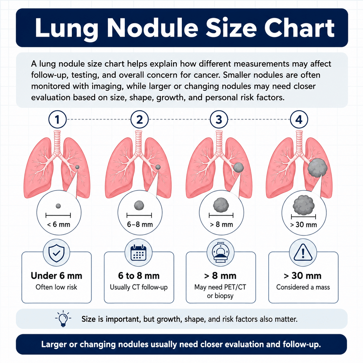
The Fleischner Society guidelines, which are commonly used for incidental lung nodules found on CT scans, base follow-up decisions partly on nodule size. Common size groups include nodules smaller than 6 millimeters, nodules 6 to 8 millimeters, and nodules larger than 8 millimeters.
Note: These size cutoffs help doctors decide whether a nodule can be watched, whether a CT scan should be repeated, or whether additional testing should be considered.
Understanding Millimeters and Centimeters
Lung nodules are usually measured in millimeters. This can be confusing because most people do not think in millimeters every day.
Here is a simple way to understand the measurements:
- 1 millimeter is very small, about the thickness of a credit card
- 5 millimeters is about the size of a small pea
- 10 millimeters is 1 centimeter
- 20 millimeters is 2 centimeters
- 30 millimeters is 3 centimeters
A nodule that measures 6 millimeters is less than one-fourth of an inch. A nodule that measures 10 millimeters is about 1 centimeter, or a little less than half an inch.
Even small differences can matter. A nodule that grows from 5 millimeters to 8 millimeters may still sound small, but that change can be important because it may show growth.
Lung Nodule Size Chart
The chart below gives a general overview of lung nodule sizes and what they may mean. This is not a diagnosis and should not replace medical advice. Your doctor may recommend a different plan based on your personal situation.
Less Than 6 Millimeters
A lung nodule smaller than 6 millimeters is considered very small. Many nodules in this size range are benign, especially in people who have a low risk of lung cancer.
For low-risk people, a very small solid nodule may not require routine follow-up. In higher-risk people, such as those with a significant smoking history, the doctor may recommend a follow-up CT scan to make sure the nodule is stable.
Small nodules can be caused by old infections, tiny scars, inflammation, or other harmless changes. Because they are so small, they are often too difficult to biopsy accurately.
6 to 8 Millimeters
A nodule in the 6 to 8 millimeter range is still small, but it usually deserves more attention than a tiny nodule. Doctors may recommend a follow-up CT scan to see whether it changes over time.
The timing of the follow-up depends on the person’s risk factors and the nodule’s appearance. A smooth, stable nodule in a low-risk person may be handled differently than an irregular nodule in a person with a long smoking history.
A major review reported that the probability of cancer is less than 1% for nodules smaller than 6 millimeters and about 1% to 2% for nodules measuring 6 to 8 millimeters.
These numbers are helpful, but they are only general estimates. Your actual risk may be higher or lower depending on other details.
Larger Than 8 Millimeters
A nodule larger than 8 millimeters is more likely to need additional evaluation. This does not mean it is cancer, but the chance is high enough that doctors often consider more than routine monitoring.
Possible next steps may include:
- A repeat CT scan in a shorter time period
- PET/CT scan
- Biopsy
- Referral to a pulmonologist
- Referral to a thoracic surgeon
The American Academy of Family Physicians summarizes that nodules larger than 8 millimeters may be evaluated with CT follow-up, PET/CT, biopsy, or removal depending on the risk level and clinical situation.
A larger nodule may still be benign, especially if it has benign calcification, fat, or long-term stability. But because larger nodules are more concerning, they usually require a clear plan.
10 Millimeters or 1 Centimeter
A 10 millimeter lung nodule is the same as a 1 centimeter nodule. This size often gets more attention because it is large enough for doctors to consider additional testing depending on its appearance and risk factors.
A 1 centimeter nodule may be watched with CT scans if it looks low risk, but a suspicious nodule may need PET imaging, biopsy, or specialist evaluation.
The decision depends on whether the nodule is solid or ground glass, whether it has smooth or irregular edges, whether it has grown, and whether the person has risk factors such as smoking or prior cancer.
20 Millimeters or 2 Centimeters
A 20 millimeter nodule is 2 centimeters. At this size, doctors usually take a closer look. A 2 centimeter nodule has a higher chance of being cancer than a tiny nodule, although benign causes are still possible.
Testing may include PET/CT, biopsy, or surgical consultation. If the nodule has been stable for a long time, that may be reassuring. If it is new or growing, further evaluation is usually needed.
30 Millimeters or 3 Centimeters
A 30 millimeter spot is 3 centimeters. This is the upper size limit for what is usually called a nodule. Larger than this, it is commonly called a lung mass.
A lung mass is more concerning than a small nodule and usually requires prompt evaluation. This may involve imaging, biopsy, and referral to a specialist.
Again, not every large spot is cancer. Infections, inflammatory disease, and benign tumors can sometimes be large. But a mass should not be ignored.
Lung Nodule Size Chart by Risk Level
A simple way to think about lung nodule size is by general risk category.
- A nodule smaller than 6 millimeters is often low risk, especially if it is smooth and found in someone without major lung cancer risk factors.
- A nodule between 6 and 8 millimeters is usually considered an intermediate finding that may need follow-up imaging.
- A nodule larger than 8 millimeters usually deserves closer evaluation, especially if it is new, growing, irregular, or found in someone with a history of smoking or cancer.
Note: This is only a general guide. A very small nodule with suspicious features may need follow-up, while a larger nodule with clearly benign features may be less concerning.
Size Is Important, But It Is Not Everything
It is natural to focus on the number in the radiology report. Many people search for “4 mm lung nodule,” “7 mm lung nodule,” or “1 cm lung nodule” because they want to know whether that specific size is dangerous.
But doctors do not make decisions based on size alone. A lung nodule is evaluated by combining several details.
Shape
Smooth, round nodules are often less concerning. Irregular, lobulated, or spiculated nodules may be more suspicious.
A spiculated nodule has tiny lines or points extending outward from the edge. This can sometimes be seen with lung cancer, but it can also occur with scarring or inflammation.
Density
A nodule may be described as solid, ground glass, or part-solid.
- A solid nodule looks dense throughout.
- A ground glass nodule looks hazy, like frosted glass.
- A part-solid nodule has both hazy and solid areas.
Note: Part-solid nodules can be more concerning than pure ground glass nodules, especially if the solid part grows over time.
Growth
Growth is one of the most important signs doctors watch for. A nodule that stays the same size over time is often less concerning than one that grows.
However, some types of lung cancer grow slowly. This is especially true for some ground glass and part-solid nodules. That is why follow-up may continue for several years in certain cases.
Location
Nodules in the upper lobes of the lungs may raise more concern, especially in people with a smoking history. Location alone does not diagnose cancer, but it adds to the overall risk assessment.
Number of Nodules
Some people have one nodule, while others have several. Multiple small nodules may be related to old infections, inflammation, or other noncancerous causes. In some cases, multiple nodules can be related to cancer or spread from another cancer.
Doctors look at the pattern, size, and appearance of all nodules, not just the largest one.
Calcification
Some nodules contain calcium. Certain patterns of calcification can suggest that a nodule is benign, especially if it is related to an old healed infection.
Not all calcification is reassuring, so the pattern matters.
Fat Within the Nodule
Fat inside a nodule can suggest a benign growth called a hamartoma. This type of nodule is usually not cancer.
What Causes Small Lung Nodules?
Small lung nodules can have many causes. The most common causes are not cancer.
Possible causes include:
- Old infections
- Fungal infections
- Tuberculosis exposure
- Scar tissue
- Inflammation
- Rheumatoid nodules
- Sarcoidosis
- Benign tumors
- Early lung cancer
- Cancer spread from another part of the body
In some regions, fungal infections are a common cause of benign nodules. These infections may leave behind small scars or granulomas even after the person has recovered.
A granuloma is a small area of inflammation that can form when the immune system walls off an infection or irritant. Granulomas are often benign.
What Size Lung Nodule Is Concerning?
There is no single size at which a lung nodule becomes definitely dangerous. Instead, risk increases gradually as size increases.
In general:
- Nodules smaller than 6 millimeters are often low risk
- Nodules 6 to 8 millimeters may need follow-up CT
- Nodules larger than 8 millimeters may need closer evaluation
- Nodules around 3 centimeters or larger are more concerning and may be called a mass
A nodule may be concerning even if it is small when it has suspicious features, such as spiculated edges, growth, or a part-solid appearance. A nodule may be less concerning even if it is larger when it has benign features, such as certain calcification patterns, fat, or long-term stability.
What Size Lung Nodule Needs a Biopsy?
There is no automatic biopsy size. Many people assume that once a nodule reaches a certain size, it must be biopsied. In reality, biopsy decisions depend on risk, location, appearance, and whether the biopsy can be done safely.
A biopsy may be considered when:
- The nodule is larger than 8 millimeters
- The nodule is growing
- The nodule has suspicious borders
- The nodule has a solid component
- PET scan results are concerning
- The person has a high risk of lung cancer
- The diagnosis would change treatment decisions
A biopsy may not be recommended right away when:
- The nodule is very small
- The nodule looks benign
- The nodule has been stable for a long time
- The biopsy would be difficult or risky
- Follow-up CT is a safer first step
Note: Very small nodules can be hard to biopsy accurately. If the needle misses the target, the result may be unclear. This is one reason doctors may choose follow-up imaging instead of immediate biopsy.
What Size Lung Nodule Needs a PET Scan?
PET scans are often more useful for larger nodules, especially those 8 millimeters or larger. Very small nodules may be too small for a PET scan to evaluate reliably.
A PET scan looks for increased metabolic activity. Cancer cells often use more energy than normal cells, but infection and inflammation can also show activity. This means a PET scan can be helpful, but it cannot always give a definite answer.
A PET scan may be recommended when a nodule is large enough and has features that raise concern. It may also be used before biopsy or surgery to help guide the next step.
What Size Lung Nodule Needs Surgery?
Surgery is not based on size alone. A larger or suspicious nodule may be removed if doctors believe the chance of cancer is high or if a biopsy is not likely to provide a clear answer.
Surgery may also be considered when a biopsy confirms cancer or when the nodule is highly suspicious and the person is healthy enough for an operation.
The type of surgery depends on the size and location of the nodule, lung function, and whether cancer is present. Options may include removing a small wedge of lung tissue, a segment, or a lobe.
Solid vs Ground Glass Nodule Size
The meaning of size can be different depending on whether the nodule is solid or ground glass.
Solid Nodules
Solid nodules are dense throughout. Many follow-up guidelines use the size categories of less than 6 millimeters, 6 to 8 millimeters, and greater than 8 millimeters.
Solid nodules that do not grow over time are often less concerning. If a solid nodule is stable for a long enough period, the doctor may decide that no further follow-up is needed.
Ground Glass Nodules
Ground glass nodules are hazy rather than fully solid. These can be caused by infection, inflammation, scarring, or slow-growing abnormal cells.
Ground glass nodules may require longer follow-up because some grow slowly. The Fleischner guidance increased the total follow-up period for persistent subsolid nodules to 5 years because of slower growth.
Part-Solid Nodules
Part-solid nodules have both ground glass and solid areas. The size of the solid portion is especially important. A growing solid component can raise concern and may lead to further testing.
Why Old Imaging Is So Helpful
If a lung nodule is found, one of the most useful things your doctor can do is compare it with older scans.
Old imaging can answer important questions:
- Was the nodule there before?
- Has it grown?
- Has it changed shape?
- Has it become more solid?
- Are there new nodules?
- Has it stayed stable for years?
Note: A nodule that has been unchanged for a long time is usually less concerning than one that is new or growing. If you had prior imaging at another hospital, clinic, or imaging center, tell your healthcare provider. The actual images are often needed, not just the written report.
Common Examples by Nodule Size
3 mm Lung Nodule
A 3 millimeter nodule is very small. Many nodules this size are benign and may not need follow-up in low-risk people. In higher-risk people, the doctor may recommend a CT scan later to make sure it has not changed. A 3 millimeter nodule is usually too small for biopsy or PET scan.
4 mm Lung Nodule
A 4 millimeter nodule is also very small. It is often found incidentally. The follow-up plan depends on risk factors and appearance. If the person has no smoking history and the nodule looks smooth, the doctor may not recommend routine follow-up. If the person has a higher risk, a repeat CT may be considered.
5 mm Lung Nodule
A 5 millimeter nodule is still in the very small range. The risk of cancer is generally low, but follow-up may depend on the person’s risk level and whether the nodule has suspicious features.
6 mm Lung Nodule
A 6 millimeter nodule is often where follow-up becomes more likely. Doctors may recommend a repeat CT scan to see whether it is stable. The timing depends on risk factors and imaging appearance.
7 mm Lung Nodule
A 7 millimeter nodule commonly requires follow-up imaging. It is still small, but large enough that doctors usually want to make sure it does not grow.
8 mm Lung Nodule
An 8 millimeter nodule is an important threshold. Nodules larger than 8 millimeters often need more detailed evaluation. Depending on the situation, this may include CT follow-up, PET/CT, biopsy, or referral to a specialist.
10 mm Lung Nodule
A 10 millimeter nodule is 1 centimeter. At this size, doctors often look closely at the nodule’s shape, density, and growth pattern. Some 1 centimeter nodules are benign, but others need further testing.
15 mm Lung Nodule
A 15 millimeter nodule is 1.5 centimeters. This size often requires a more complete evaluation, especially if it is new, growing, or irregular.
20 mm Lung Nodule
A 20 millimeter nodule is 2 centimeters. This is considered a larger nodule. It may need PET/CT, biopsy, or surgical evaluation depending on the risk level.
30 mm Lung Nodule
A 30 millimeter nodule is 3 centimeters. This is at the upper limit of the nodule category. A spot larger than this is usually called a lung mass and often requires prompt evaluation.
How Doctors Decide the Follow-Up Plan
Doctors use size along with the person’s risk factors to decide what happens next.
Low-Risk Patients
A low-risk person may be younger, have never smoked, have no prior cancer, and have no major exposures such as asbestos or radon. In low-risk people, small nodules may need little or no follow-up, especially if they look benign.
High-Risk Patients
A higher-risk person may be older, have a history of smoking, have prior cancer, or have significant exposure to radon, asbestos, or other lung cancer risks. In higher-risk people, doctors may recommend closer follow-up, even for smaller nodules.
Imaging Features
The CT appearance can change the plan. A small nodule with smooth edges may be watched less aggressively than one with irregular or spiculated edges. A part-solid nodule may need longer follow-up than a solid nodule of similar size.
What Happens During Follow-Up?
Follow-up usually involves repeat CT scans. The goal is to see whether the nodule stays the same, gets smaller, disappears, or grows.
- If the nodule disappears, it may have been caused by infection or inflammation.
- If it stays the same, that may be reassuring, although some nodules still need continued monitoring.
- If it grows, becomes more solid, or develops suspicious features, more testing may be needed.
Your doctor should tell you:
- When the next scan should happen
- What changes they are watching for
- Whether you need a specialist
- Whether PET scan or biopsy may be needed
- When follow-up can stop
Symptoms to Watch For
Most small lung nodules do not cause symptoms. Many people feel completely normal. However, symptoms should be discussed with a healthcare provider, especially if they are new, persistent, or worsening.
Symptoms that may need medical attention include:
- Persistent cough
- Coughing up blood
- New or worsening shortness of breath
- Chest pain
- Unexplained weight loss
- Hoarseness
- Repeated pneumonia
- Fatigue that does not improve
- Fever that does not go away
Note: Seek emergency care for severe trouble breathing, severe chest pain, fainting, blue lips, or coughing up a large amount of blood.
Lung Cancer Screening and Nodule Size
Some lung nodules are found during lung cancer screening. Screening is done with low-dose CT in people who meet certain risk criteria, usually based on age and smoking history.
Lung cancer screening can find small nodules before symptoms occur. Many nodules found during screening are not cancer, but some need follow-up based on size and appearance.
Screening programs may use a system called Lung-RADS, which helps categorize findings and recommend next steps. This system is different from the Fleischner guidelines used for many incidental nodules, but both consider size and imaging features.
If you have a smoking history, ask your healthcare provider whether lung cancer screening is appropriate for you.
Questions to Ask Your Doctor
If you are told you have a lung nodule, ask your healthcare provider to explain the finding in plain language.
Helpful questions include:
- How large is the nodule in millimeters?
- Is it solid, ground glass, or part-solid?
- Does it have smooth or irregular edges?
- Where is it located?
- Was it present on older scans?
- Has it grown?
- Do I need a follow-up CT scan?
- When should the next scan be done?
- Do I need a PET scan?
- Do I need a biopsy?
- Should I see a lung specialist?
- What symptoms should I report?
- When can follow-up stop?
Note: These questions can help you understand the plan and reduce uncertainty.
Why You Should Not Rely on Size Alone
A lung nodule size chart can be helpful, but it should not be used to make a diagnosis by itself. The same size nodule can mean different things in different people.
For example, a 7 millimeter smooth nodule in a young nonsmoker may be low risk. A 7 millimeter spiculated nodule in an older person with a long smoking history may be more concerning.
A 10 millimeter nodule that has been stable for many years may be less concerning than a 6 millimeter nodule that is new and growing.
Note: This is why your doctor’s interpretation matters. The number is important, but the pattern is just as important.
Final Thoughts
A lung nodule size chart can help you understand why doctors respond differently to different nodules. In general, nodules smaller than 6 millimeters are often low risk, nodules between 6 and 8 millimeters may need follow-up CT scans, and nodules larger than 8 millimeters may need closer evaluation.
Still, size is only one part of the picture. Shape, density, growth, location, smoking history, age, and prior cancer history all matter. If you have been told you have a lung nodule, the most important step is to follow your provider’s plan and keep any recommended imaging appointments.

Written by:
John Landry is a registered respiratory therapist from Memphis, TN, and has a bachelor's degree in kinesiology. He enjoys using evidence-based research to help others breathe easier and live a healthier life.
References
- Loverdos K, Fotiadis A, Kontogianni C, Iliopoulou M, Gaga M. Lung nodules: A comprehensive review on current approach and management. Ann Thorac Med. 2019.