
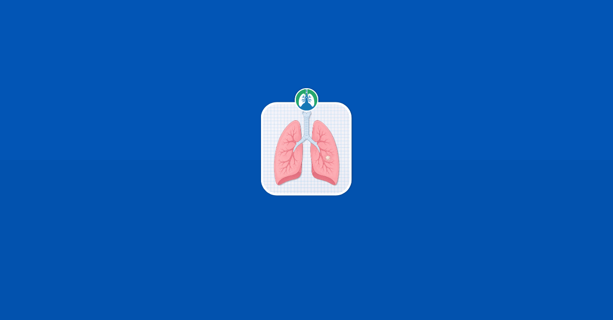
A spiculated lung nodule is a small spot in the lung that has an irregular or star-like edge on an imaging scan. The word “spiculated” can sound alarming, especially because this type of border can sometimes be linked with lung cancer.
However, a spiculated nodule is not automatically cancer. Infections, inflammation, scar tissue, and other noncancerous conditions can also cause abnormal-looking nodules.
The most important step is careful follow-up with a healthcare provider who can look at the nodule’s size, shape, location, growth, and your overall risk factors.

Download our free guide that has over 100+ of the best tips for healthy lungs.
What Is a Lung Nodule?
A lung nodule is a small area of abnormal tissue seen on an imaging test, such as a chest X-ray or CT scan. It may also be called a pulmonary nodule. Many people first learn they have one after having imaging done for another reason, such as a cough, chest pain, pneumonia, injury, or routine lung cancer screening.
Lung nodules are common. In many cases, they are small, harmless, and caused by old infections or areas of scarring. Most lung nodules found on CT scans are not cancer and are more often related to old infections, scar tissue, or other causes.
A nodule is usually described as a spot or small mass in the lung. Doctors often use the word “nodule” when the spot is 3 centimeters or smaller. A spot larger than that may be called a lung mass, which generally requires a more urgent evaluation.
A lung nodule can vary in:
- Size
- Shape
- Border appearance
- Density
- Location
- Growth pattern
- Number of nodules present
Note: Some nodules are smooth and round. Others are irregular. Some are solid, while others look partly cloudy or hazy on CT imaging. These details matter because they help doctors estimate how likely the nodule is to be benign or cancerous.
What Does “Spiculated” Mean?
“Spiculated” means the border of the lung nodule looks irregular, sharp, or star-like. Instead of having a smooth, rounded edge, the nodule may appear to have tiny lines or spikes extending outward into the surrounding lung tissue.
These pointed edges are called spicules. On a CT scan, they can make the nodule look like it is pulling on nearby tissue or spreading into the lung around it.
A simple way to picture it is this:
- A smooth nodule may look more like a small marble.
- A spiculated nodule may look more like a small burr, star, or sea urchin, with uneven points around the outside.
This does not prove cancer, but it does make the nodule more suspicious. Spiculated or irregular margins are among the imaging features that doctors consider when estimating cancer risk. Irregular or spiculated margins are among the factors that may increase concern when evaluating pulmonary nodules.

Why Spiculated Nodules Get More Attention
Doctors pay close attention to spiculated lung nodules because cancerous nodules can sometimes grow in an irregular pattern. As a cancer grows, it may interact with nearby lung tissue, blood vessels, lymph channels, or scar-like tissue. This can create a pulling or radiating appearance around the edge of the nodule.
That said, spiculation is only one piece of the picture. A healthcare provider does not look at the border alone. They also consider the nodule’s size, whether it has changed over time, whether it is solid or subsolid, where it is located, and whether the person has risk factors such as smoking history or older age.
A small spiculated nodule that has not changed for years may be handled differently than a new spiculated nodule that is larger and growing. The same imaging word can mean different things depending on the full situation.
Is a Spiculated Lung Nodule Always Cancer?
No. A spiculated lung nodule is not always cancer.
This is one of the most important points to understand. Spiculation increases concern, but it does not provide a diagnosis by itself. Imaging can suggest that a nodule is suspicious, but a definite diagnosis often requires more information, such as comparison with old scans, follow-up imaging, PET scan findings, or a biopsy.
Noncancerous causes of spiculated or irregular lung nodules may include:
- Old lung infections
- Fungal infections
- Tuberculosis or prior healed infection
- Inflammatory lung disease
- Scar tissue
- Rheumatoid nodules
- Granulomas
- Organizing pneumonia
- Areas of prior injury or irritation
Cancerous causes may include:
- Early lung cancer
- Cancer that started elsewhere and spread to the lung
- Less common lung tumors
Note: The challenge is that benign and cancerous nodules can sometimes look similar on imaging. This is why doctors use a step-by-step process rather than making assumptions from one word in a report.
Why Lung Nodules Are Often Found by Accident
Many lung nodules do not cause symptoms. They are often too small to affect breathing or cause pain. Because of this, a person may feel completely normal and still have a nodule show up on a scan.
Common reasons a nodule may be found include:
- A CT scan done after a respiratory infection
- A scan after chest pain or injury
- Lung cancer screening
- Imaging before surgery
- A scan done to check another condition
- Follow-up imaging after pneumonia
- A chest X-ray that leads to a CT scan
Note: CT scans can detect much smaller nodules than regular chest X-rays. As CT scans have become more common, more people are being told they have lung nodules. This can be stressful, but finding a nodule early can also be helpful. If it is harmless, monitoring can provide reassurance. If it turns out to be cancer, early detection may allow more treatment options.
How Doctors Estimate the Risk of Cancer
A spiculated border is important, but it is not the only factor. Doctors estimate risk by combining imaging features with personal health factors.
Size of the Nodule
Size is one of the most important factors. In general, larger nodules are more concerning than smaller ones. A tiny nodule may have a very low risk, while a larger nodule, especially one over 8 millimeters, may need closer evaluation.
Nodules greater than 8 millimeters are often evaluated with short-term follow-up CT, PET/CT, biopsy, or removal depending on the full clinical picture. Size alone does not diagnose cancer. Some small nodules are cancerous, and some large nodules are benign. Still, size helps guide what happens next.
Growth Over Time
One of the most useful pieces of information is whether the nodule is growing. If a nodule has been stable for a long time, that can be reassuring, especially for solid nodules. If it is growing, changing shape, or becoming more solid, doctors usually become more concerned.
This is why old imaging is so valuable. A CT scan from several years ago can help determine whether the nodule is new or has been present and stable for a long time.
Shape and Border
A smooth, round nodule is often less suspicious than one with irregular, lobulated, or spiculated edges. A spiculated border suggests that the nodule is interacting with surrounding lung tissue in a way that deserves closer attention. However, inflammation and scarring can also cause irregular edges. This is why the border is important but not final.
Density
Doctors also look at how the nodule appears inside.
A nodule may be described as:
- Solid
- Ground-glass
- Part-solid
- Calcified
- Fat-containing
Note: Certain types of calcification can suggest a benign cause. Fat inside a nodule can sometimes point toward a benign tumor called a hamartoma. Ground-glass and part-solid nodules can require special follow-up because some slow-growing lung cancers may appear this way.
Location in the Lung
Nodules in the upper lobes of the lungs may carry more concern, especially in people with a history of smoking. Location is not enough to diagnose cancer, but it adds to the overall risk estimate.
Smoking History
Smoking is one of the strongest risk factors for lung cancer. A spiculated nodule in a person with a long smoking history is usually taken more seriously than the same finding in a younger person who has never smoked.
Doctors may ask:
- Do you currently smoke?
- Did you smoke in the past?
- How many years did you smoke?
- How many packs per day did you smoke?
- When did you quit?
Note: This information helps estimate risk and decide on the next step.
Age
The chance that a lung nodule is cancer generally increases with age. Nodules in younger adults are often benign, especially when there are no other risk factors. In older adults, doctors may recommend closer follow-up.
Personal or Family History of Cancer
A history of lung cancer or another type of cancer may change the evaluation. Some cancers can spread to the lungs, and people who have had cancer before may need a more careful workup. A family history of lung cancer may also be considered, especially when combined with smoking or other exposures.
Environmental and Occupational Exposures
Certain exposures may increase lung cancer risk, including:
- Radon
- Asbestos
- Diesel exhaust
- Silica dust
- Secondhand smoke
- Some workplace chemicals
Note: Your healthcare provider may ask about your work history, home environment, and exposure risks.
Common Causes of a Spiculated Lung Nodule
A spiculated lung nodule can have several possible causes. Some are benign, while others are cancerous. The goal of evaluation is to narrow down the most likely explanation.
Lung Cancer
A spiculated lung nodule may represent early lung cancer. This is why the finding needs attention. When lung cancer is found as a small nodule, it may be at an earlier stage than cancer found after symptoms develop.
Possible signs that increase concern include:
- Larger size
- Growth over time
- Spiculated or irregular margins
- Upper-lobe location
- Part-solid appearance
- New nodule compared with older scans
- Smoking history
- Older age
Note: Even when these features are present, more testing is needed before a diagnosis can be made.
Old Infection
Prior infections can leave behind small scars or granulomas in the lung. A granuloma is a small area of inflammation that can form after the immune system walls off an infection or irritant.
Some granulomas are caused by fungal infections, which are more common in certain regions. Others may be linked to prior tuberculosis exposure or other infections. These nodules may be benign but can sometimes look irregular.
Inflammation
Inflammatory conditions can cause lung nodules. Examples include rheumatoid arthritis, sarcoidosis, and certain immune-related lung diseases. Inflammatory nodules may come and go, remain stable, or appear with other lung findings. Blood tests, symptoms, medical history, and imaging patterns may help guide the diagnosis.
Scar Tissue
Scar tissue can form after infection, surgery, radiation, injury, or long-term irritation. Scar-related nodules may look irregular because scar tissue can pull on nearby lung structures.
Sometimes it is difficult to tell whether an irregular area is only scarring or something more concerning. Comparison with older imaging can be especially helpful.
Organizing Pneumonia
Organizing pneumonia is a type of lung inflammation that can appear as nodules or patchy areas on imaging. It is not the same as typical bacterial pneumonia, although it can occur after infection, medication reactions, autoimmune disease, or unknown causes.
Because it can mimic cancer on imaging, doctors may need follow-up scans, medication trials, or biopsy in some cases.
Symptoms of a Spiculated Lung Nodule
Most small lung nodules do not cause symptoms. A person may have no cough, no shortness of breath, and no pain.
When symptoms are present, they may be caused by the nodule, the condition that caused the nodule, or an unrelated lung problem.
Possible symptoms may include:
- Persistent cough
- Coughing up blood
- Shortness of breath
- Chest discomfort
- Wheezing
- Repeated lung infections
- Unexplained weight loss
- Fatigue
- Hoarseness
Note: These symptoms do not automatically mean cancer, but they should be discussed with a healthcare provider. Coughing up blood, worsening shortness of breath, chest pain, or unexplained weight loss should be taken seriously and evaluated promptly.
What the Radiology Report May Say
Seeing medical terms in a radiology report can be confusing. A report may include words that sound alarming but are meant to describe what the radiologist sees.
The report may mention:
- “Spiculated nodule”
- “Irregular margins”
- “Suspicious morphology”
- “Solid pulmonary nodule”
- “Part-solid nodule”
- “Ground-glass opacity”
- “Upper lobe nodule”
- “Recommend follow-up CT”
- “Recommend PET/CT”
- “Tissue sampling may be considered”
Note: These phrases help your doctor decide the next step. They do not always mean that cancer has been diagnosed. A radiology report is not the same as a final diagnosis. It is one part of the evaluation.
Tests Used to Evaluate a Spiculated Lung Nodule
The next test depends on the nodule’s size, appearance, risk level, and whether prior imaging is available.
Comparing With Older Scans
One of the first steps is often to compare the current scan with any older chest imaging. This may include previous CT scans, chest X-rays, or scans of the abdomen that included the lower lungs.
If the nodule was present years ago and has not changed, that may reduce concern. If it is new or growing, more testing may be needed. Patients can help by telling their doctor where previous imaging was done. Sometimes scans from another hospital or imaging center need to be requested.
Follow-Up CT Scan
A follow-up CT scan may be recommended to see whether the nodule changes over time. The timing can vary. Some nodules are checked in a few months, while others may be followed over a longer period.
Follow-up CT is commonly used when the nodule is not clearly harmless but does not immediately require biopsy. The goal is to watch for growth or change. A low-dose CT technique may be used in some cases to reduce radiation exposure while still providing useful images.
PET Scan
A PET scan, often combined with CT, can help evaluate whether a nodule is metabolically active. Cancer cells often use more energy than normal cells, so they may show increased activity on PET imaging.
However, PET scans are not perfect. Infections and inflammation can also “light up,” leading to false positives. Small cancers or slow-growing cancers may not show strong PET activity, leading to false negatives. PET/CT is often considered for nodules that are large enough for the test to be useful, especially nodules 8 millimeters or larger.
Biopsy
A biopsy means removing a small sample of tissue so it can be examined under a microscope. This is often the only way to know for sure whether a suspicious nodule is cancer.
A biopsy may be done in different ways, depending on the nodule’s location:
- CT-guided needle biopsy through the chest wall
- Bronchoscopy, where a thin tube is passed through the mouth or nose into the airways
- Robotic or navigational bronchoscopy for harder-to-reach nodules
- Surgical biopsy
CT-guided needle biopsy is one method used when a suspicious area is deep in the body and CT guidance can help direct the needle.
A biopsy has benefits and risks. Possible risks include bleeding, infection, or a collapsed lung, also called pneumothorax. Your doctor will weigh these risks against the concern for cancer.
Surgical Removal
In some cases, a nodule may be removed surgically, especially if the chance of cancer is high or if a biopsy is difficult to perform safely.
Surgery may involve removing:
- A small wedge of lung tissue
- A lung segment
- A lobe of the lung
Note: The best approach depends on the nodule, the person’s lung function, overall health, and whether cancer is suspected or confirmed.
Why Some Nodules Are Watched Instead of Biopsied Right Away
It can feel unsettling to be told that a suspicious nodule will be monitored instead of removed immediately. However, observation can be appropriate in certain situations.
Doctors may recommend follow-up rather than immediate biopsy when:
- The nodule is very small
- The biopsy would be difficult or risky
- The risk of cancer is low to moderate
- The nodule could represent infection or inflammation
- More time is needed to see whether it grows
- The person has health conditions that make invasive testing risky
A biopsy is not always simple. Very small nodules can be hard to sample accurately. Some nodules are located near blood vessels or deep in the lung. In these cases, the safest plan may be careful imaging follow-up.
Note: The goal is not to ignore the nodule. The goal is to choose the safest and most useful next step.
Questions to Ask Your Doctor
If your report says “spiculated lung nodule,” it is reasonable to ask clear questions. Many people feel anxious after reading the report, and having a plan can help.
Helpful questions include:
- How large is the nodule?
- Is it solid, ground-glass, or part-solid?
- Where is it located?
- Was it present on any older scans?
- Has it grown or changed?
- What features make it concerning?
- What features are reassuring?
- What is my estimated risk level?
- Do I need a follow-up CT scan, PET scan, biopsy, or specialist referral?
- When should the next step happen?
- Should I see a pulmonologist, thoracic surgeon, or lung nodule clinic?
- What symptoms should I report right away?
- Should I be screened for lung cancer in the future?
Note: You do not need to understand every technical detail, but you should understand the plan.
When to Seek Medical Attention Promptly
A spiculated lung nodule should be followed carefully, but some symptoms require quicker attention.
Contact a healthcare provider promptly if you develop:
- Coughing up blood
- New or worsening shortness of breath
- Chest pain that is persistent or severe
- Unexplained weight loss
- Ongoing fever
- Repeated pneumonia in the same area
- A cough that does not improve
- New hoarseness that persists
- Severe fatigue with other concerning symptoms
Note: Seek emergency care for severe trouble breathing, severe chest pain, fainting, blue lips, or coughing up a large amount of blood.
Lung Cancer Screening and Spiculated Nodules
Some lung nodules are found through lung cancer screening. Screening is meant to detect lung cancer early in people at higher risk before symptoms appear.
In the United States, lung cancer screening is generally recommended for certain adults based on age and smoking history. Guidelines can change, so people should ask their healthcare provider whether they qualify.
Screening is done with low-dose CT, not a regular chest X-ray. Low-dose CT can find small nodules earlier, but it can also find nodules that are not cancer. This is why follow-up guidelines are important.
If a spiculated nodule is found during screening, the radiology report may recommend a specific follow-up category or next step. This may involve repeat imaging, PET/CT, or referral to a specialist.
What Happens If the Nodule Is Cancer?
If testing shows that the nodule is cancer, the next step is staging. Staging means finding out how large the cancer is, whether lymph nodes are involved, and whether it has spread elsewhere.
Early-stage lung cancer may be treated with surgery, radiation, or other targeted approaches. More advanced cancer may require a combination of treatments, such as chemotherapy, immunotherapy, targeted therapy, radiation, or surgery in selected cases.
The exact plan depends on:
- Cancer type
- Stage
- Genetic or molecular markers
- Lung function
- Overall health
- Patient preferences
- Whether the cancer has spread
Note: A small spiculated nodule that turns out to be cancer may still be treatable, especially if found early. This is one reason follow-up is important.
What Happens If the Nodule Is Benign?
If the nodule is determined to be benign, treatment may not be needed. In some cases, doctors may continue to monitor it for a period of time to make sure it stays stable.
If the nodule is caused by infection, inflammation, or another condition, treatment may focus on that cause. For example, an infection may require medication, while an inflammatory condition may require evaluation by a specialist.
Some benign nodules remain in the lung permanently as scar tissue. They may continue to show up on future scans. Keeping a record of prior imaging can help prevent unnecessary worry later.
Living With Uncertainty During Follow-Up
Waiting for follow-up imaging or test results can be stressful. Many people immediately fear the worst when they hear the word “spiculated.” That reaction is understandable, but it is important to remember that the word describes appearance, not a final diagnosis.
During this period, it may help to focus on what can be controlled:
- Keep follow-up appointments
- Ask for clear timelines
- Obtain old imaging records if possible
- Write down questions before visits
- Avoid repeatedly searching worst-case scenarios online
- Share concerns with your healthcare team
- Stop smoking if you currently smoke
- Stay up to date with recommended medical care
Note: If anxiety becomes overwhelming, tell your healthcare provider. Emotional stress is a real part of the nodule evaluation process.
Why Follow-Up Should Not Be Delayed
Although many lung nodules are benign, a spiculated nodule deserves timely follow-up. Delaying recommended imaging or testing can make it harder to catch important changes early.
Follow-up is especially important if the nodule is:
- Larger than 8 millimeters
- New
- Growing
- Spiculated or irregular
- Part-solid
- Located in an upper lobe
- Found in a person with a smoking history
- Found in a person with prior cancer
Note: This does not mean panic is needed. It means the finding should be taken seriously and managed with a clear plan.
How to Prepare for Your Appointment
Before seeing your doctor or specialist, gather useful information. This can make the visit more productive.
Bring or prepare:
- A copy of the radiology report
- The date and location of the scan
- Any older chest imaging records
- A list of symptoms
- Smoking history
- Occupational exposure history
- Family history of lung cancer
- Prior cancer history
- Current medications
- Questions you want answered
Note: If your imaging was done at a different facility, ask whether the actual images can be sent to your doctor, not just the written report. Radiologists and specialists often need to review the images directly.
How Doctors Decide Between CT, PET Scan, and Biopsy
There is no single next step that fits everyone. The decision is based on risk.
- A lower-risk nodule may be watched with CT scans.
- An intermediate-risk nodule may need PET/CT, biopsy, or close follow-up.
- A high-risk nodule may need biopsy or surgical evaluation.
Doctors may also use risk calculators that combine age, smoking history, nodule size, spiculation, location, and other features. These tools do not give perfect answers, but they can support decision-making.
The choice also depends on the patient’s health. For example, someone with severe lung disease may not be a good candidate for surgery, so the team may choose a different diagnostic or treatment pathway.
Can Lifestyle Changes Make a Spiculated Nodule Go Away?
Lifestyle changes cannot reliably make a spiculated lung nodule disappear. However, healthy changes can support overall lung health and reduce future risk.
The most important step for people who smoke is to stop smoking. Quitting smoking lowers the risk of lung cancer and other lung diseases over time. It also improves heart health and surgical recovery if a procedure is ever needed.
Other helpful steps include:
- Avoid secondhand smoke
- Test your home for radon if recommended in your area
- Use protective equipment around dust or chemicals
- Stay current with vaccines recommended by your provider
- Manage chronic lung conditions
- Keep follow-up imaging appointments
- Report new symptoms promptly
Note: These steps do not replace medical evaluation, but they can support better long-term health.
Common Misunderstandings About Spiculated Lung Nodules
“Spiculated Means Cancer”
This is not always true. Spiculation is suspicious, but it is not a diagnosis. Some benign conditions can create irregular or spiculated-looking nodules.
“No Symptoms Means It Cannot Be Serious”
Many small lung cancers do not cause symptoms at first. A lack of symptoms is reassuring in some ways, but it does not eliminate the need for follow-up.
“A PET Scan Gives a Definite Answer”
PET scans are useful, but they are not perfect. Infection and inflammation can cause increased activity, while some cancers may show little activity. PET results must be interpreted with the full clinical picture.
“A Biopsy Is Always the First Step”
Not always. Some nodules are too small or difficult to biopsy safely. In other cases, follow-up imaging may be the better first step.
“If It Is Small, It Does Not Matter”
Small nodules are often benign, but size is only one factor. A small nodule with suspicious features may still need follow-up.
Final Thoughts
A spiculated lung nodule is an imaging finding that should be taken seriously, but it is not the same as a cancer diagnosis. The term means the nodule has an irregular or star-like border, which can raise concern and may require closer evaluation. The next step depends on the nodule’s size, appearance, growth, location, and your personal risk factors.
Some spiculated nodules are cancerous, while others are caused by infection, inflammation, or scar tissue. The best approach is timely follow-up, clear communication with your healthcare provider, and a plan that matches your individual situation.

Written by:
John Landry is a registered respiratory therapist from Memphis, TN, and has a bachelor's degree in kinesiology. He enjoys using evidence-based research to help others breathe easier and live a healthier life.
References
- Qiu S, Sun J, Zhou T, Gao G, He Z, Liang T. Spiculation Sign Recognition in a Pulmonary Nodule Based on Spiking Neural P Systems. Biomed Res Int. 2020.