

Thoracic imaging plays an important role in evaluating respiratory and cardiovascular disorders. It allows clinicians to examine the lungs, pleural spaces, airways, mediastinum, heart, diaphragm, chest wall, and the position of medical devices.
The chest x-ray remains the most commonly used imaging study because it is fast, inexpensive, and widely available. However, computed tomography, magnetic resonance imaging, ultrasound, positron emission tomography, angiography, and ventilation-perfusion scanning provide additional information when plain radiographs are not sufficient.
Understanding basic imaging principles helps clinicians recognize common abnormalities and interpret them in the proper clinical context.

What is Thoracic Imaging?
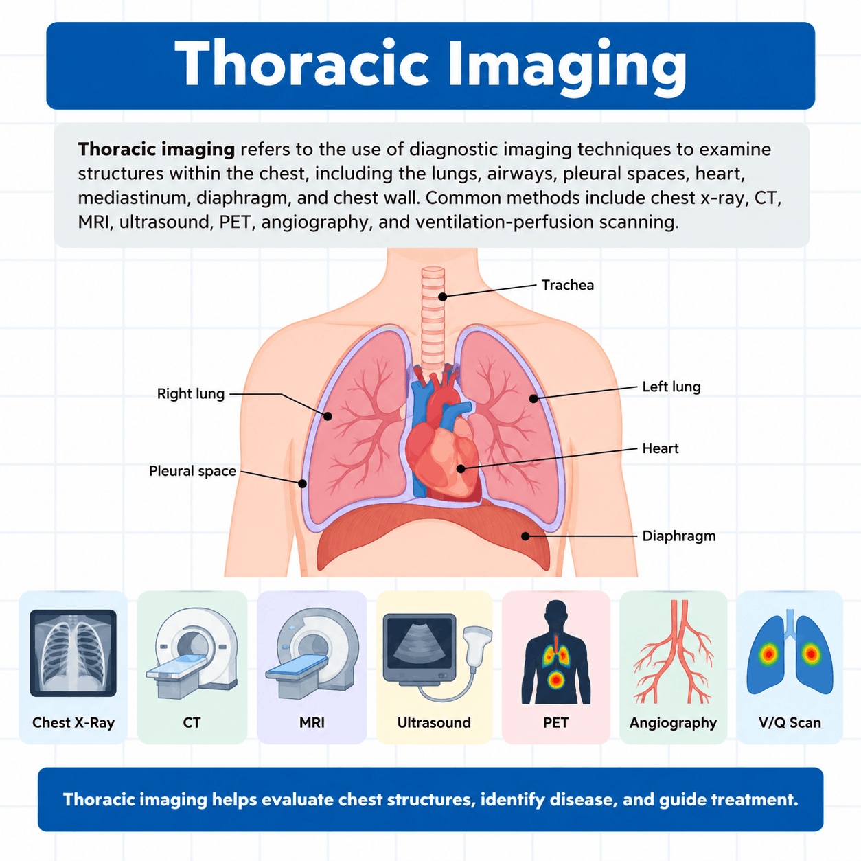
Thoracic imaging refers to the use of diagnostic imaging techniques to examine structures within the chest. It is commonly used to evaluate the lungs, airways, pleural spaces, heart, mediastinum, diaphragm, chest wall, and the position of medical devices such as endotracheal tubes, chest tubes, and vascular catheters.
The chest x-ray is the most frequently used form of thoracic imaging because it is fast, widely available, and useful for identifying conditions such as pneumonia, atelectasis, pneumothorax, pleural effusion, pulmonary edema, and hyperinflation.
Other imaging methods provide additional detail when needed. Computed tomography offers detailed cross-sectional views of the chest, while MRI is useful for selected soft-tissue and cardiovascular abnormalities. Ultrasound is valuable for detecting pleural fluid and pneumothorax at the bedside. PET imaging, angiography, and ventilation-perfusion scanning can provide metabolic, vascular, or functional information.
Thoracic imaging findings should always be interpreted together with the patient’s clinical condition.
Chest X-Ray Fundamentals
A chest x-ray is produced when x-rays pass through the thorax and strike an image receptor. Different tissues absorb different amounts of radiation, creating the shades seen on the final image.
Four major densities are commonly recognized:
- Air
- Fat
- Soft tissue or water
- Bone
Air absorbs relatively little radiation and therefore appears dark, or radiolucent. Because normal lungs contain large amounts of air, the lung fields normally appear relatively dark.
Fat is slightly denser than air and appears somewhat lighter. Soft tissues and fluid absorb more radiation and therefore appear as varying shades of gray or white. Bone absorbs a large amount of radiation and appears bright white.
Metallic objects are even more radiopaque than bone. As a result, coins, surgical wires, pacemaker components, and radiopaque portions of tubes and catheters are usually easy to identify. Understanding these differences in density is essential because most abnormalities are recognized by changes in the normal pattern of radiolucency and radiopacity.
The Silhouette Sign
Normal anatomical borders can be seen when two structures with different densities are positioned next to each other. For example, the soft-tissue density of the heart is easily distinguished from the surrounding air-filled lungs.
When two structures of similar density come into direct contact, however, the border between them may disappear. This is known as the silhouette sign.
The silhouette sign can help determine the location of pulmonary disease. For example, the right middle lobe lies adjacent to the right heart border. If the right middle lobe becomes filled with inflammatory material, fluid, or collapsed lung tissue, the normal right heart border may become obscured.
Loss of the right heart border therefore suggests disease involving the right middle lobe. Similar relationships can help localize abnormalities elsewhere in the chest.
Indications for Thoracic Imaging
Chest imaging may be obtained for many respiratory and cardiovascular conditions. Common outpatient indications include persistent cough, hemoptysis, unexplained dyspnea, fever with sputum production, severe chest pain, and evaluation following certain positive infectious disease screenings.
In hospitalized or critically ill patients, imaging may be obtained when there is:
- Sudden respiratory deterioration
- New or worsening hypoxemia
- Acute chest pain
- Suspected pneumothorax
- Significant change in mechanical ventilator pressures
- Placement of an endotracheal or tracheostomy tube
- Placement of a central venous catheter
- Placement of a pulmonary artery catheter
- Placement of a chest tube
- Concern regarding the location of a feeding tube
- Suspected complications following an invasive thoracic procedure
Note: Imaging should always be interpreted together with the patient’s symptoms, examination findings, laboratory results, oxygenation, ventilation, and response to treatment.
A Systematic Approach to Chest X-Ray Interpretation
A consistent method of chest x-ray interpretation reduces the likelihood of missing important findings. A dramatic abnormality can easily distract the observer from a second, more subtle problem.
For example, a large pulmonary infiltrate may be obvious, while a small pneumothorax, misplaced catheter, rib fracture, or free air under the diaphragm may be overlooked.
A systematic review should begin with verification of the patient’s identity and the date of the image. The observer should then determine the projection and assess image quality before examining the anatomy.
Important structures to assess include:
- Bones and soft tissues
- Trachea
- Mediastinum
- Heart
- Hilar structures
- Diaphragms
- Costophrenic angles
- Pleural spaces
- Lung fields
- Upper abdominal structures
- Tubes, lines, and other devices
Note: Comparison with previous imaging is also valuable because a change over time may be more important than a single isolated finding.
Evaluating Image Quality
A technically poor chest x-ray can create the appearance of disease or hide a true abnormality. Rotation, poor inspiration, incorrect positioning, and improper exposure must therefore be recognized before making clinical conclusions.
Rotation
The patient should be positioned symmetrically when a frontal chest radiograph is obtained. The medial ends of the clavicles should appear approximately equal distances from the spinous processes.
Significant rotation can distort the appearance of the trachea, mediastinum, hila, heart, and pulmonary vessels. It may falsely suggest mediastinal widening or abnormal hilar positioning.
Inspiration
Adequate inspiration is essential because low lung volumes can make the lungs appear excessively dense and can exaggerate the apparent size of the heart.
On a well-inspired PA chest x-ray, approximately six anterior ribs or ten posterior ribs are generally visible above the diaphragm. Poor inspiration may mimic pulmonary disease by producing crowding of vascular markings and apparent bibasilar opacities.
Position
The observer should determine whether the image is PA, AP, lateral, decubitus, or another projection.
The PA projection is preferred when possible because it provides a more accurate representation of heart size. AP images are often obtained at the bedside in critically ill patients. Because the heart lies farther from the detector in this position, it appears magnified.
Exposure
An adequately exposed radiograph should allow the intervertebral spaces to be faintly visible through the cardiac silhouette.
An underexposed image appears too white and may exaggerate pulmonary markings or obscure deeper structures. An overexposed image appears too dark and may cause subtle infiltrates or pneumothorax to be missed.
PA, AP, and Lateral Chest Radiographs
The standard outpatient chest examination generally includes PA and lateral projections.
PA Projection
During a PA chest x-ray, the x-ray beam passes from the posterior chest to the anterior chest. The patient’s anterior chest is positioned close to the detector. This arrangement minimizes magnification of the heart and provides a more accurate estimate of cardiac size.
AP Projection
The AP projection is frequently used for portable bedside imaging. The x-ray beam passes from the anterior chest toward the posterior chest. AP radiographs are common in intensive care units because many critically ill patients cannot stand or travel safely to the radiology department.
Because of the geometry of the projection, the heart often appears larger than it actually is. Apparent cardiomegaly on an AP film should therefore be interpreted cautiously.
Lateral Projection
A lateral chest x-ray provides an additional perspective and helps localize abnormalities that overlap on a frontal image.
Lesions positioned behind the heart or diaphragm may be difficult to identify on the PA view alone. The lateral projection can help determine whether an abnormality is located in the upper or lower lobe and whether it lies anteriorly or posteriorly.
Radiolucency and Radiopacity
Radiolucent structures appear darker because they allow more x-rays to pass through. Radiopaque structures appear lighter because they absorb more radiation.
Conditions that may increase radiolucency include:
- Emphysema
- Pneumothorax
- Pneumomediastinum
- Pneumopericardium
- Subcutaneous emphysema
- Pulmonary interstitial emphysema
Conditions that may increase radiopacity include:
- Atelectasis
- Pneumonia
- Pulmonary edema
- Pleural effusion
- Interstitial lung disease
- Tumors
- Pulmonary hemorrhage
- Calcification
Note: The pattern, distribution, and associated findings help determine the most likely explanation for an abnormal density.
Pneumothorax
A pneumothorax occurs when air enters the pleural space and separates the visceral pleura from the chest wall. On an upright chest radiograph, a thin visceral pleural line may be visible. Beyond this line, toward the chest wall, normal pulmonary vascular markings are absent. The affected region therefore appears unusually radiolucent.
A pneumothorax can be more difficult to detect in a supine patient because pleural air tends to move anteriorly and inferiorly instead of collecting at the apex.
One important finding on a supine film is the deep sulcus sign. The costophrenic region becomes abnormally deep and radiolucent. Bullae and skin folds can occasionally mimic pneumothorax. If normal pulmonary markings extend beyond a suspicious line, a true pneumothorax is less likely.
Tension Pneumothorax
A tension pneumothorax occurs when pleural air accumulates under pressure. As pressure increases, the affected lung collapses and mediastinal structures may shift away from the involved side. The diaphragm on the affected side may become depressed. This differs from atelectasis, in which loss of lung volume generally pulls mediastinal structures toward the abnormal side.
A useful general rule is:
Volume loss pulls structures toward the problem, while increased pressure or a large space-occupying process pushes structures away.
Tension pneumothorax is a clinical emergency. Imaging may support the diagnosis, but treatment should not be delayed when the clinical findings strongly indicate the condition.
Pleural Effusion
A pleural effusion is an abnormal accumulation of fluid within the pleural space. On an upright chest radiograph, pleural fluid typically collects at the lung bases. One of the earliest findings is blunting of the costophrenic angle.
As the amount of fluid increases, a curved upper border called the meniscus may become visible. Large effusions can compress the adjacent lung and push the mediastinum away from the affected side.
A lateral decubitus film can help determine whether pleural fluid moves freely. Free-flowing fluid shifts toward the dependent side, while loculated fluid may remain fixed. Thoracic ultrasound is especially useful for confirming pleural fluid and selecting a safe location for thoracentesis.
Empyema
An empyema is infected fluid within the pleural space. CT imaging is particularly useful in evaluating suspected empyema because it can demonstrate localized pleural collections, pleural thickening, and surrounding inflammatory changes.
Gas may sometimes be visible within an infected pleural collection. Because empyema may resemble other pleural abnormalities on plain radiographs, CT and ultrasound frequently provide additional diagnostic information.
Atelectasis
Atelectasis refers to loss of lung volume caused by collapse of alveoli. It may involve a small segment, a lobe, or an entire lung. Common causes include mucus plugging, foreign-body obstruction, tumors, hypoventilation, and postoperative shallow breathing.
Radiographic findings may include:
- Increased opacity
- Elevated hemidiaphragm
- Narrowed intercostal spaces
- Displacement of pulmonary fissures
- Hilar displacement
- Mediastinal shift toward the affected side
Note: Upper-lobe atelectasis may pull the hilum upward. The presence of volume loss is one of the main features that helps distinguish atelectasis from consolidation.
Consolidation
Consolidation occurs when normal alveolar air is replaced by fluid, inflammatory material, blood, cells, or aspirated material. The involved portion of the lung becomes more radiopaque.
Pneumonia is a common cause of consolidation, although pulmonary edema, hemorrhage, and aspiration can produce similar appearances.
Consolidation may affect a segment, an entire lobe, or multiple regions of the lung. Unlike atelectasis, consolidation does not necessarily produce major loss of lung volume.
Air Bronchograms
An air bronchogram occurs when air remains within the bronchi while the surrounding alveoli become filled with material. The bronchi then appear as dark branching structures within a lighter region of consolidated lung.
Air bronchograms commonly occur with:
- Pneumonia
- Pulmonary edema
- Pulmonary hemorrhage
- Other forms of alveolar filling
Note: Their presence suggests that the abnormality is located within the lung parenchyma rather than within the pleural space.
Pulmonary Edema
Pulmonary edema occurs when fluid accumulates within the pulmonary interstitial tissues and alveoli.
Cardiogenic pulmonary edema most commonly results from elevated pulmonary vascular pressure caused by left-sided heart dysfunction. An early radiographic sign is cephalization, or redistribution of pulmonary blood flow toward the upper lobes.
Normally, pulmonary vessels are larger in the lower lungs because of gravity. As pulmonary venous pressure rises, upper-lobe vessels become more prominent.
Additional findings can include:
- Increased hilar vascular markings
- Perihilar haze
- Peribronchial cuffing
- Kerley B lines
- Bilateral alveolar opacities
- Pleural effusions
- Cardiomegaly
Note: Severe alveolar edema may produce a central perihilar distribution sometimes described as a batwing appearance.
Cardiogenic Pulmonary Edema vs. ARDS
Cardiogenic pulmonary edema and acute respiratory distress syndrome may both produce bilateral pulmonary opacities.
Cardiogenic edema is more likely to be associated with cardiomegaly, cephalization, vascular congestion, Kerley B lines, and pleural effusions. ARDS may produce diffuse or patchy bilateral infiltrates without cardiomegaly or classic findings of elevated pulmonary venous pressure.
A normal-sized heart in a patient with extensive bilateral opacities may support a noncardiogenic process, but imaging findings must always be interpreted with the patient’s clinical presentation.
Interstitial Lung Disease
The pulmonary interstitium forms the supporting framework surrounding alveoli, blood vessels, and airways. Under normal circumstances, most interstitial structures are too thin to be seen on a conventional chest x-ray.
Interstitial disease can produce:
- Reticular opacities
- Linear opacities
- Nodules
- Septal thickening
- Cysts
- Honeycombing
Note: Many interstitial disorders produce diffuse bilateral abnormalities. High-resolution CT is considerably more sensitive than chest radiography for identifying the pattern, distribution, and severity of interstitial lung disease.
Emphysema and Hyperinflation
Emphysema is associated with destruction of alveolar walls, reduced pulmonary vascular markings, and hyperinflation.
Common imaging findings include:
- Flattened diaphragms
- Increased lung volume
- Increased radiolucency
- Widened intercostal spaces
- Reduced vascular markings
- More horizontal ribs
- Increased retrosternal air space
- Relatively narrow or elongated cardiac silhouette
Note: These findings may also occur to varying degrees in other obstructive conditions associated with significant air trapping. CT provides much greater detail than chest x-ray when evaluating the distribution and severity of emphysema.
Mediastinal and Tracheal Shifts
The position of the trachea and mediastinum provides important information regarding changes in lung volume and intrathoracic pressure.
Conditions associated with volume loss tend to pull the mediastinum toward the affected side. Examples include:
- Atelectasis
- Pulmonary fibrosis
- Surgical lung resection
Conditions that increase pressure or occupy a large amount of space may push the mediastinum away from the affected side. Examples include:
- Tension pneumothorax
- Large pleural effusion
- Large thoracic mass
Note: Patient rotation must always be considered because poor positioning can falsely suggest mediastinal displacement.
The Diaphragm
The hemidiaphragms normally have a domed appearance and form sharp costophrenic angles with the chest wall.
An elevated hemidiaphragm may occur with:
- Atelectasis
- Pulmonary fibrosis
- Phrenic nerve dysfunction
- Abdominal enlargement
- Hepatomegaly on the right side
A depressed or flattened diaphragm may occur with:
- Emphysema
- Severe asthma
- Other causes of hyperinflation
- Tension pneumothorax
Note: Free air beneath the diaphragm can suggest perforation of an abdominal organ and should not be confused with the normal gastric air bubble.
Heart Size
Heart size is most accurately assessed on a properly obtained PA chest radiograph. The cardiothoracic ratio compares the transverse diameter of the heart with the internal transverse diameter of the thorax.
In adults, a cardiothoracic ratio below approximately 0.50 is generally considered normal. A larger ratio may indicate cardiomegaly.
Heart size should not be judged with the same confidence on an AP portable film because magnification may falsely enlarge the cardiac silhouette.
Solitary Pulmonary Nodules
A solitary pulmonary nodule is a rounded opacity measuring less than 3 cm in diameter and surrounded by aerated lung. An opacity measuring 3 cm or greater is generally classified as a pulmonary mass.
CT is preferred for evaluating pulmonary nodules because it provides detailed information about:
- Size
- Shape
- Margins
- Internal density
- Calcification
- Fat content
- Relationship to surrounding structures
Fat within a nodule may suggest a hamartoma, while certain central calcification patterns may support a benign granuloma. Irregular or spiculated margins raise greater concern for malignancy.
PET-CT may provide additional information by assessing metabolic activity, although increased tracer uptake is not specific for cancer.
Mediastinal Abnormalities
The mediastinum contains the heart, great vessels, trachea, esophagus, lymphatic structures, thymus, and other tissues. Mediastinal abnormalities are often classified according to location.
Anterior mediastinal lesions may include:
- Thymic abnormalities
- Lymphoma
- Thyroid or parathyroid masses
- Teratomas
Middle mediastinal abnormalities may include:
- Lymphadenopathy
- Bronchogenic cysts
- Vascular abnormalities
- Tracheal or esophageal lesions
Posterior mediastinal abnormalities may include:
- Neurogenic tumors
- Descending aortic aneurysms
- Certain hernias
Note: CT is generally preferred for evaluating mediastinal masses because it clearly demonstrates their location and relationship to adjacent structures.
Pneumomediastinum and Other Air Leaks
Pneumomediastinum refers to free air within the mediastinum. It may result from airway injury, esophageal perforation, trauma, barotrauma, or air tracking from another anatomical region. Radiographically, air may outline mediastinal structures and extend into the soft tissues of the neck.
Subcutaneous emphysema occurs when air collects within the soft tissues. It may produce streaky or bubbly radiolucent areas beneath the skin.
Pneumopericardium refers to air within the pericardial space. Pulmonary interstitial emphysema occurs when air dissects through the interstitial tissues of the lung and is especially associated with mechanically ventilated premature infants.
Endotracheal Tube Position
Chest radiography is commonly used to verify the position of an endotracheal tube.
In an adult with the neck in a neutral position, the tube tip should generally lie several centimeters above the carina. A commonly used target is approximately 4 to 6 cm above the carina, although acceptable positioning may vary slightly depending on patient anatomy and head position.
A tube positioned too high may be accidentally displaced through the vocal cords. A tube advanced too far commonly enters the right mainstem bronchus because the right main bronchus is wider and more vertically oriented.
Note: Right mainstem intubation may cause inadequate ventilation of the left lung, leading to atelectasis and hypoxemia.
Tracheostomy Tubes and Chest Tubes
The distal end of a tracheostomy tube should remain centered within the trachea and above the carina. Chest tubes are placed within the pleural space to remove air, fluid, blood, or infected material.
Tubes intended primarily for pneumothorax drainage are generally directed toward regions where pleural air is expected to collect, whereas tubes intended primarily for fluid drainage are directed toward dependent pleural regions. Chest imaging can help assess both position and treatment response.
Central Venous and Pulmonary Artery Catheters
Central venous catheters may be evaluated radiographically to confirm tip position and identify complications such as pneumothorax.
Pulmonary artery catheters are advanced through the right side of the heart and into the pulmonary circulation. The distal tip should ideally remain within a proximal pulmonary artery.
Excessive advancement into a peripheral pulmonary artery can increase the risk of vessel obstruction, infarction, pseudoaneurysm, or pulmonary artery rupture. Imaging is therefore useful when catheter position is uncertain or when complications are suspected.
Intra-Aortic Balloon Pump Position
The intra-aortic balloon pump contains a radiopaque marker that allows its location to be evaluated on chest x-ray. The upper portion of the device should remain in the proximal descending thoracic aorta, just distal to the origin of the left subclavian artery.
The carina can provide a useful radiographic reference point. A device positioned too high may interfere with blood flow to vessels supplying the head and upper extremities. A position that is too low may compromise blood flow to abdominal organs.
Neck Radiographs
Neck radiographs can be useful when upper-airway obstruction is suspected, particularly in pediatric patients.
Croup may produce narrowing of the subglottic airway, resulting in the classic steeple sign on an AP neck radiograph. Epiglottitis can produce enlargement of the epiglottis on a lateral neck radiograph, creating the thumb sign.
Radiopaque foreign bodies may also be visible. However, food, plastic, and other nonmetallic materials may not be directly seen on x-ray. In these cases, indirect findings and additional procedures such as bronchoscopy or laryngoscopy may be required.
Computed Tomography
Computed tomography (CT) provides detailed cross-sectional images of the thorax and eliminates much of the anatomical overlap seen on conventional radiographs.
Thoracic CT is useful for evaluating:
- Pulmonary nodules and masses
- Interstitial lung disease
- Emphysema
- Pleural abnormalities
- Mediastinal masses
- Chest trauma
- Abscesses
- Vascular abnormalities
- Complicated infections
Intravenous contrast may be used to distinguish vascular structures and improve visualization of blood vessels. CT angiography is particularly useful when pulmonary embolism is suspected.
CT can also generate multiplanar and three-dimensional reconstructions. Airway reconstructions may assist with procedural planning and provide virtual bronchoscopy-type views. The main disadvantage of CT compared with conventional chest radiography is greater radiation exposure.
Magnetic Resonance Imaging
Magnetic resonance imaging (MRI) uses a strong magnetic field and radiofrequency energy instead of ionizing radiation. It provides excellent soft-tissue contrast and is useful for evaluating:
- Mediastinal masses
- Major thoracic vessels
- Cardiac structures
- Aortic disease
- Chest wall abnormalities
- Selected tumors
MRI is generally less useful than CT for routine evaluation of air-filled lung tissue. The lack of ionizing radiation can be especially beneficial in selected patients, including children.
Because of the strong magnetic field, implanted devices and monitoring equipment must be evaluated for MRI compatibility.
Thoracic Ultrasound
Thoracic ultrasound has become increasingly important in critical care because it can be performed rapidly at the bedside without ionizing radiation.
Important applications include:
- Detecting pleural effusion
- Identifying pneumothorax
- Evaluating selected pulmonary abnormalities
- Assessing cardiac function
- Estimating intravascular volume
- Guiding thoracentesis
- Guiding central venous access
- Assisting with arterial and chest tube placement
Normal movement between the visceral and parietal pleura produces lung sliding. Absence of lung sliding may occur with pneumothorax, although other clinical conditions can also reduce or eliminate this finding.
B-lines appear as bright vertical artifacts extending downward from the pleural surface. Multiple B-lines may occur with interstitial or alveolar processes such as pulmonary edema. Pleural fluid typically appears as a dark or echo-free collection in a dependent region.
PET-CT
Positron emission tomography evaluates tissue metabolism using a radioactive tracer. PET-CT combines metabolic information from PET with detailed anatomical imaging from CT.
Malignant cells often demonstrate increased metabolic activity, making PET-CT useful in the evaluation and staging of suspected cancer. However, increased tracer uptake is not specific for malignancy. Infection and inflammation may also produce increased metabolic activity.
PET findings must therefore be interpreted together with CT characteristics and the patient’s clinical presentation.
Ventilation-Perfusion Scanning
A ventilation-perfusion scan compares airflow within the lungs with pulmonary blood flow.
The ventilation portion demonstrates the distribution of inhaled radioactive gas or aerosol. The perfusion portion demonstrates pulmonary blood flow after intravenous administration of a radioactive tracer.
Pulmonary embolism can produce a region that remains ventilated but receives little or no perfusion. This creates a ventilation-perfusion mismatch. V/Q scanning remains useful in selected patients, particularly when CT angiography is not appropriate.
Pulmonary Angiography
Pulmonary angiography involves injecting contrast material directly into the pulmonary circulation through a catheter. Normal pulmonary arteries fill with contrast and appear as branching vascular structures. A pulmonary embolus may appear as a filling defect or interruption of blood flow.
CT angiography has replaced conventional pulmonary angiography for many diagnostic applications, but catheter-based angiography may still be used in selected cases and can sometimes allow therapeutic intervention.
Imaging During Mechanical Ventilation
Chest imaging is particularly important in mechanically ventilated patients because both disease progression and treatment complications can alter the appearance of the thorax.
Radiography can help evaluate:
- Endotracheal tube position
- Atelectasis
- Pneumothorax
- Pulmonary edema
- Pleural effusion
- Pneumonia
- Barotrauma
- Central venous catheter position
- Pulmonary artery catheter position
- Chest tube position
A sudden increase in airway pressure, worsening hypoxemia, unequal breath sounds, or asymmetric chest movement may prompt imaging to evaluate for problems such as pneumothorax, mainstem intubation, or acute atelectasis.
Note: Clinical findings and imaging should be used together rather than interpreted independently.
Key Takeaways
Several relationships are especially important when evaluating respiratory imaging.
- Atelectasis causes volume loss and tends to pull the mediastinum toward the affected side.
- Tension pneumothorax creates increasing pressure and can push the mediastinum away from the affected side.
- Large pleural effusions may also push mediastinal structures away.
- Pleural fluid commonly produces blunting of the costophrenic angle.
- Pneumothorax produces an area without normal peripheral vascular markings.
- Emphysema commonly produces hyperinflation, flattened diaphragms, and increased radiolucency.
- Cardiogenic pulmonary edema may produce vascular congestion, cardiomegaly, Kerley B lines, pleural effusions, and bilateral alveolar opacities.
- ARDS may produce extensive bilateral infiltrates without the cardiomegaly commonly associated with cardiogenic pulmonary edema.
- Consolidation produces increased opacity and may contain air bronchograms.
- Croup may produce a steeple sign.
- Epiglottitis may produce a thumb sign.
- A deeply inserted endotracheal tube most commonly enters the right mainstem bronchus.
- The PA projection is preferred when evaluating heart size because AP portable films can artificially magnify the cardiac silhouette.
Thoracic Imaging Practice Questions
1. What is the most commonly used imaging study for evaluating the thorax?
Chest x-ray
2. Which four fundamental tissue densities can be identified on a chest radiograph?
Air, fat, soft tissue or water, and bone
3. Why do normal air-filled lungs appear relatively dark on a chest radiograph?
Air absorbs relatively little x-ray radiation, allowing more x-rays to pass through to the detector.
4. What is the silhouette sign?
The silhouette sign is the loss of a normally visible border between two adjacent structures when they have similar radiographic densities.
5. Loss of the right heart border on a chest radiograph most commonly suggests disease involving which lung lobe?
Right middle lobe
6. Which chest radiograph projection is preferred for the most accurate assessment of heart size?
Posteroanterior (PA) projection
7. Why can the heart appear artificially enlarged on an AP portable chest radiograph?
The heart is farther from the image detector and is therefore magnified.
8. Approximately how many posterior ribs should be visible above the diaphragm on a well-inspired PA chest radiograph?
Approximately 10 posterior ribs
9. How can proper patient rotation be assessed on a frontal chest radiograph?
The spinous processes should be approximately midway between the medial ends of the clavicles.
10. What appearance does an underpenetrated chest radiograph typically have?
It appears excessively white and may exaggerate pulmonary vascular markings.
11. What radiographic finding is characteristic of a pneumothorax?
A visible visceral pleural line with absence of pulmonary vascular markings beyond it toward the chest wall
12. What is the deep sulcus sign?
An abnormally deep and radiolucent costophrenic region that may indicate pneumothorax in a supine patient.
13. In which direction does the mediastinum typically shift with a tension pneumothorax?
Away from the affected side
14. In which direction does the mediastinum typically shift with significant unilateral atelectasis?
Toward the affected side
15. What chest x-ray finding commonly suggests a pleural effusion?
Blunting of the costophrenic angle
16. What is an air bronchogram?
A dark, air-filled bronchus that becomes visible against surrounding radiopaque alveolar consolidation.
17. What is cephalization on a chest radiograph?
Redistribution of pulmonary blood flow toward the upper lobes due to elevated pulmonary venous pressure.
18. What are Kerley B lines?
Short horizontal peripheral lines caused by fluid or thickening within the interlobular septa.
19. Which radiographic findings commonly accompany cardiogenic pulmonary edema?
Cardiomegaly, pulmonary vascular congestion, cephalization, Kerley B lines, bilateral alveolar opacities, and pleural effusions
20. What radiographic feature can help distinguish ARDS from cardiogenic pulmonary edema?
ARDS may produce diffuse bilateral infiltrates without cardiomegaly or the classic vascular congestion associated with cardiogenic pulmonary edema.
21. What imaging study is more sensitive than a conventional chest x-ray for evaluating interstitial lung disease?
High-resolution computed tomography (CT)
22. What chest x-ray findings are commonly associated with emphysema?
Hyperinflation, flattened diaphragms, increased radiolucency, widened intercostal spaces, and reduced pulmonary vascular markings
23. How is a solitary pulmonary nodule generally defined?
A rounded pulmonary opacity measuring less than 3 cm in diameter and surrounded by aerated lung.
24. Where should the tip of an adult endotracheal tube generally be positioned on a chest radiograph?
Several centimeters above the carina, commonly about 4 to 6 cm with the neck in a neutral position.
25. Which mainstem bronchus is most commonly entered when an endotracheal tube is advanced too far?
Right mainstem bronchus
26. What type of chest radiograph can help determine whether pleural fluid is free-flowing?
Lateral decubitus radiograph
27. What does a meniscus sign suggest on a chest radiograph?
Pleural fluid forming a curved upper border within the pleural space
28. What is an empyema?
An infected collection of fluid within the pleural space
29. Which imaging modality is especially useful for confirming pleural fluid before thoracentesis?
Thoracic ultrasound
30. What radiographic feature helps distinguish atelectasis from simple consolidation?
Evidence of lung volume loss
31. What happens to the diaphragm on the affected side of significant atelectasis?
It may become elevated.
32. What happens to the intercostal spaces in an area of marked lung volume loss?
They may become narrowed.
33. What does hilar displacement toward an abnormal lung region suggest?
Atelectasis or another process causing lung volume loss
34. What is the batwing appearance associated with?
Severe central or perihilar alveolar pulmonary edema
35. What causes peribronchial cuffing on a chest radiograph?
Fluid accumulation around the bronchi
36. What is the pulmonary interstitium?
The supporting tissue framework surrounding the alveoli, airways, and blood vessels
37. What does honeycombing on thoracic imaging usually suggest?
Advanced chronic interstitial lung disease or fibrosis
38. Which imaging modality is preferred for detailed characterization of a pulmonary nodule?
Computed tomography (CT)
39. What nodule characteristic can suggest a pulmonary hamartoma?
The presence of fat within the nodule
40. What type of nodule margin raises greater concern for malignancy?
An irregular or spiculated margin
41. What is the primary advantage of PET-CT when evaluating a pulmonary nodule?
It provides information about the metabolic activity of the lesion.
42. Why can increased tracer uptake on PET-CT not be considered diagnostic of cancer by itself?
Inflammation and infection can also produce increased metabolic activity.
43. Which imaging study is generally preferred for evaluating most mediastinal masses?
Computed tomography (CT)
44. What is pneumomediastinum?
The presence of free air within the mediastinum
45. What can cause pneumomediastinum?
Airway injury, esophageal perforation, chest trauma, or air dissecting into the mediastinum from another region
46. What is subcutaneous emphysema?
The presence of air within the soft tissues beneath the skin
47. What is pneumopericardium?
The presence of air within the pericardial space surrounding the heart
48. Which pediatric condition is associated with the steeple sign on an AP neck radiograph?
Croup
49. Which pediatric condition is associated with the thumb sign on a lateral neck radiograph?
Epiglottitis
50. What is a major limitation of conventional chest radiography when evaluating some aspirated foreign bodies?
Nonmetallic materials such as food and some plastics may not be directly visible.
51. What is the main advantage of CT compared with a standard chest x-ray?
CT provides detailed cross-sectional images that reduce overlap of thoracic structures.
52. Why is intravenous contrast commonly used during thoracic CT imaging?
To improve visualization of blood vessels and distinguish vascular structures from surrounding tissues.
53. Which CT technique is especially useful for evaluating suspected pulmonary embolism?
CT pulmonary angiography
54. What is one major disadvantage of CT compared with conventional chest radiography?
CT exposes the patient to a higher dose of ionizing radiation.
55. What type of energy does MRI use to produce images?
A strong magnetic field and radiofrequency energy
56. Why is MRI generally less useful than CT for routine imaging of the lungs?
Air-filled lung tissue produces relatively poor MRI detail.
57. Which thoracic structures are especially well evaluated with MRI?
The mediastinum, heart, great vessels, chest wall, and selected vascular structures
58. What is an important safety consideration before performing MRI?
The patient and all equipment must be evaluated for compatibility with the magnetic field.
59. What is one major advantage of MRI over CT?
MRI does not use ionizing radiation.
60. What is the purpose of a ventilation-perfusion scan?
To compare the distribution of ventilation with pulmonary blood flow.
61. What V/Q scan pattern is classically associated with pulmonary embolism?
A perfusion defect in an area where ventilation remains present
62. What is pulmonary angiography?
An imaging procedure in which contrast is injected into the pulmonary circulation to visualize pulmonary blood vessels.
63. How can a pulmonary embolus appear during pulmonary angiography?
As a filling defect or interruption in contrast flow within a pulmonary artery
64. What is the cardiothoracic ratio?
The ratio of the transverse width of the heart to the internal transverse width of the chest.
65. What cardiothoracic ratio is generally considered normal in an adult on a PA chest radiograph?
Less than approximately 0.50
66. Why should heart size be interpreted cautiously on an AP portable chest radiograph?
AP positioning magnifies the cardiac silhouette and may falsely suggest cardiomegaly.
67. What is the normal appearance of the costophrenic angles on a chest radiograph?
They should appear sharp and well defined.
68. What does bilateral depression and flattening of the hemidiaphragms commonly suggest?
Lung hyperinflation, such as that seen in emphysema or severe obstructive lung disease.
69. What can unilateral elevation of a hemidiaphragm indicate?
Atelectasis, pulmonary fibrosis, phrenic nerve dysfunction, or an abdominal process
70. What is pulmonary interstitial emphysema?
Air dissecting through the interstitial tissues of the lung.
71. In which patient population is pulmonary interstitial emphysema especially associated with mechanical ventilation?
Premature infants with respiratory distress syndrome.
72. What ultrasound finding normally occurs as the visceral and parietal pleura move against one another during breathing?
Lung sliding
73. What ultrasound finding may raise suspicion for pneumothorax?
Absence of normal lung sliding
74. What are B-lines on thoracic ultrasound?
Bright vertical artifacts extending from the pleural line that may be associated with interstitial or alveolar abnormalities.
75. What conditions can produce multiple B-lines on thoracic ultrasound?
Pulmonary edema and other interstitial or alveolar processes, including some cases of pneumonia
76. What imaging modality can detect relatively small pleural effusions at the bedside?
Thoracic ultrasound
77. Why is ultrasound guidance useful during thoracentesis?
It helps identify pleural fluid and a safer needle path for drainage.
78. Why are veins often easier to identify than arteries during ultrasound-guided vascular access?
Veins are generally more compressible than arteries.
79. What type of imaging information does PET primarily provide?
Physiologic and metabolic activity
80. Why can a lateral chest radiograph be helpful when a lesion is difficult to localize on a frontal image?
It provides a second perspective that helps determine the lesion’s anterior, posterior, and lobar location.
81. What structure should normally remain near the midline on a properly positioned chest radiograph?
The trachea
82. Why must patient rotation be considered before diagnosing tracheal or mediastinal displacement?
Rotation can create a false appearance of abnormal displacement.
83. What does absence of pulmonary vascular markings beyond a pleural line suggest?
Pneumothorax
84. What finding can help distinguish a skin fold from a true pneumothorax?
Normal lung markings can often be seen beyond a skin fold.
85. What is the usual appearance of pleural fluid on thoracic ultrasound?
A dark or echo-free collection, usually in a dependent region.
86. What may happen to the mediastinum when a very large pleural effusion develops?
It may shift away from the affected side.
87. What type of material can replace alveolar air and cause consolidation?
Fluid, inflammatory material, blood, cells, or aspirated material
88. What is one common postoperative cause of lower-lobe atelectasis?
Shallow breathing due to pain following thoracic or abdominal surgery
89. What does increased retrosternal air space on a lateral chest x-ray suggest?
Hyperinflation, commonly associated with emphysema
90. What may happen to the ribs in severe pulmonary hyperinflation?
They may appear more horizontal.
91. What does a relatively narrow and elongated cardiac silhouette sometimes indicate?
Severe lung hyperinflation, such as that seen in emphysema
92. What is the general size cutoff between a pulmonary nodule and a pulmonary mass?
A lesion less than 3 cm is generally called a nodule, while a lesion 3 cm or larger is generally called a mass.
93. What type of calcification pattern within a pulmonary nodule may suggest a benign granuloma?
Characteristic central calcification
94. What is the purpose of three-dimensional CT airway reconstruction?
To provide detailed spatial views of the airways for evaluation and procedural planning.
95. What is virtual bronchoscopy?
A CT-based three-dimensional reconstruction that creates an internal view of the airways similar to bronchoscopy.
96. Where should a pulmonary artery catheter ideally terminate?
Within the proximal right or left pulmonary artery
97. Why is persistent peripheral wedging of a pulmonary artery catheter dangerous?
It can obstruct pulmonary blood flow and contribute to infarction, pseudoaneurysm, or pulmonary artery rupture.
98. Where should the upper marker of an intra-aortic balloon pump generally be positioned?
In the proximal descending thoracic aorta just distal to the origin of the left subclavian artery.
99. Why can an intra-aortic balloon pump positioned too low be dangerous?
It may interfere with blood flow to abdominal organs.
100. Why should thoracic imaging findings always be correlated with the patient’s clinical condition?
Because imaging alone does not provide the complete diagnosis and must be interpreted with symptoms, examination findings, laboratory data, and treatment response.
Final Thoughts
Thoracic imaging provides valuable information about pulmonary disease, pleural abnormalities, cardiovascular changes, airway problems, and the position of medical devices. Chest radiography remains the primary imaging method because it is fast, accessible, and useful in both routine and critical-care settings.
Accurate interpretation begins with assessing image quality and then following a consistent review of the thoracic structures. CT, MRI, ultrasound, PET-CT, angiography, and V/Q scanning provide additional structural or functional information when needed. Imaging findings should always be correlated with the patient’s history, physical examination, respiratory status, laboratory data, and response to treatment.

Written by:
John Landry is a registered respiratory therapist from Memphis, TN, and has a bachelor's degree in kinesiology. He enjoys using evidence-based research to help others breathe easier and live a healthier life.
References
- Nishino M, Schiebler ML. Advances in Thoracic Imaging: Key Developments in the Past Decade and Future Directions. Radiology. 2023.