When it comes to detecting lung cancer, early and accurate diagnosis can mean the difference between effective treatment and missed opportunities for intervention. Computed tomography (CT) scans are one of the most widely used imaging tools in this process, offering detailed cross-sectional views of the lungs that go beyond the capabilities of a standard chest X-ray.
But just how accurate are CT scans in identifying lung cancer?
While they are highly sensitive in detecting lung nodules and abnormalities, questions remain about their ability to distinguish between benign and malignant growths. This article explores the accuracy of CT scans for lung cancer and the factors that influence their reliability.
Download our free guide that has over 100+ of the best tips for healthy lungs.
How Accurate is a CT Scan for Lung Cancer?
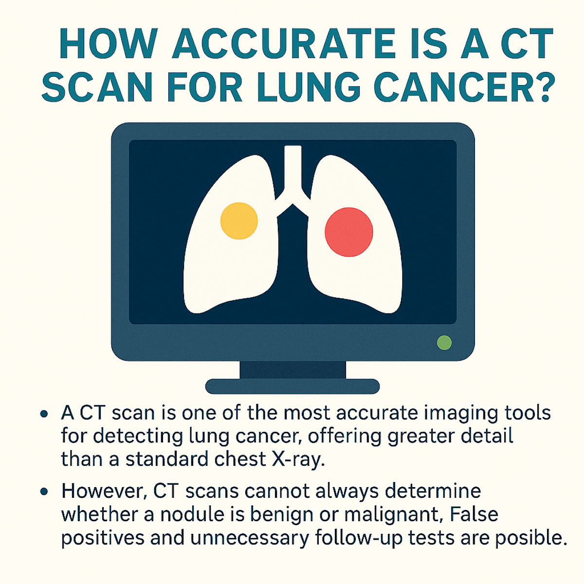
A CT scan is one of the most accurate imaging tools for detecting lung cancer, offering greater detail than a standard chest X-ray. It can identify small lung nodules and abnormalities with high sensitivity, often spotting issues at earlier stages.
However, while CT scans are excellent at detecting suspicious growths, they cannot always determine whether a nodule is benign or malignant. False positives and unnecessary follow-up tests are possible, especially in patients with infections or scarring. Overall, CT scans are highly reliable for lung cancer screening, particularly when combined with patient history and additional diagnostic tests.

CT Scan Accuracy for Detecting Lung Cancer
CT scans provide detailed images of lung tissues, making them valuable for identifying tumors and abnormalities. The accuracy depends on several measurable factors, such as sensitivity, specificity, and comparison to other techniques. Performance also varies by patient and technical variables.
Sensitivity and Specificity Rates
CT scans typically show sensitivity rates ranging from 85% to 95% for detecting lung cancer, meaning they correctly identify most true positive cases. Specificity, which measures how well the test avoids false positives, generally falls between 75% and 90%.
High sensitivity helps detect small nodules early, especially with low-dose CT used in screening programs. However, specificity is limited by benign nodules being mistaken for cancer. False positives may lead to additional testing or invasive procedures.
Note: These rates may differ based on the size and location of tumors. Larger lesions are easier to detect, increasing sensitivity, while smaller lesions under 5mm are harder to classify accurately.
Comparison With Other Imaging Techniques
Compared to chest X-rays, CT scans offer significantly higher accuracy in identifying lung cancer. While X-rays detect larger masses, CT provides 3D views that enable detection of smaller nodules.
Positron Emission Tomography (PET) combined with CT improves specificity by highlighting metabolically active tumors. PET/CT can reduce false positives but is less sensitive in very small lesions than CT alone.
Magnetic Resonance Imaging (MRI) is less commonly used for lung cancer detection but excels in assessing chest wall or mediastinal invasion once cancer is identified by CT.
Factors Impacting Test Performance
Image quality depends on the CT scanner’s technology, such as slice thickness and reconstruction algorithms. Thin-slice CT (1-1.25 mm) increases detection accuracy for small nodules.
Patient factors, including body size, breathing motion, and prior lung conditions like scarring or infections, can obscure results and reduce accuracy.
Radiologist experience also plays a role. Specialized thoracic radiologists tend to produce more accurate readings. Finally, adherence to standardized scanning protocols ensures consistent sensitivity and specificity.
Role of CT Scans in Early Lung Cancer Detection
CT scans are crucial for identifying lung abnormalities, especially in their earliest stages. They can detect very small nodules, target high-risk groups for screening, and influence patient treatment pathways.
Detection of Small Nodules
CT scans offer high-resolution images that can reveal lung nodules as small as 2-3 millimeters. This capability allows clinicians to identify suspicious growths before symptoms develop.
Early nodules detected on CT require careful evaluation to distinguish benign from malignant lesions. The scan’s sensitivity enables follow-up monitoring and timely biopsy if changes occur.
Note: Because chest X-rays often miss nodules smaller than 7-10 millimeters, CT provides a significant advantage in early lung cancer detection.
Screening in High-Risk Populations
Low-dose CT (LDCT) is the preferred method for screening individuals with heavy smoking histories or occupational exposures. It reduces radiation exposure compared to standard CT scans, making repeated screenings safer.
Screening programs using LDCT have shown a reduction in lung cancer mortality by detecting tumors at earlier, more treatable stages. Guidelines typically recommend annual LDCT scans for adults aged 50-80 with a 20 pack-year smoking history.
Note: The success of screening depends on adherence to protocol and proper interpretation of scan results alongside clinical risk factors.
Impact on Patient Outcomes
Early detection through CT scans improves surgical options and survival rates for lung cancer patients. Tumors found at a small size or limited stage often qualify for curative surgery rather than palliative care.
CT scans aid in staging cancer accurately, guiding treatment decisions such as surgery, chemotherapy, or radiation. Early-stage identification correlates with higher five-year survival rates.
Note: Regular imaging follow-up after initial detection helps monitor treatment response and detect recurrence early, further influencing long-term outcomes.
Limitations of CT Imaging for Lung Cancer
CT scans have value but also distinct limitations affecting accuracy. These include errors in identifying cancer presence and difficulties in interpreting the nature of detected nodules.
False Positives and False Negatives
CT scans can produce false positives by identifying benign lung nodules or infections as cancer. This often leads to unnecessary biopsies or treatments, causing patient anxiety and additional medical costs.
False negatives also occur when early-stage or very small tumors are missed. Nodules hidden by surrounding tissues or those with low contrast can escape detection. This sometimes delays diagnosis and treatment.
Note: The rate of false findings varies by population risk, CT technology, and radiologist experience. Low-dose CT reduces radiation but may slightly increase false negatives.
Challenges With Nodule Characterization
CT imaging struggles to definitively distinguish malignant nodules from benign ones based solely on appearance. Features like size, shape, and growth rate provide clues but are not conclusive.
Some benign lesions mimic cancer on CT, such as granulomas or scar tissue. Conversely, certain cancers may appear less suspicious and cause diagnostic uncertainty.
Further testing, such as PET scans, biopsy, or serial imaging, is often required to clarify the nature of the nodule. Reliance on CT alone limits confident decision-making in many cases.
Recent Advances in CT Technology for Lung Cancer
CT scanning for lung cancer detection has improved with enhanced imaging techniques and computational analysis. These advances allow for better identification of small nodules and offer improved safety profiles during screening.
Low-Dose CT Scans
Low-dose CT (LDCT) scans have become a standard for lung cancer screening, especially in high-risk patients like smokers. LDCT reduces radiation exposure by up to 90% compared to conventional CT while maintaining sufficient image quality to detect nodules as small as 4 mm.
This reduction in dose lowers the risk of radiation-induced harm during repeated screenings. Studies have shown LDCT screening decreases lung cancer mortality by detecting tumors earlier, before symptoms occur. Many screening programs now prefer LDCT because it balances safety and diagnostic effectiveness.
Protocols for LDCT focus on optimizing scanner settings, including lower tube voltage and current. These adjustments maintain clarity for tumor identification while minimizing dose, making LDCT vital in ongoing lung cancer surveillance.
Clinical Guidelines for CT Scan Use in Lung Cancer Assessment
CT scans play a critical role in lung cancer detection and management. Clear protocols determine who should be screened and how to proceed after uncertain or suspicious results.
Recommended Screening Protocols
The U.S. Preventive Services Task Force (USPSTF) recommends annual low-dose CT (LDCT) scans for adults aged 50-80 with a 20 pack-year smoking history who currently smoke or have quit within the past 15 years.
Screening should continue only if the individual is healthy enough for potential lung surgery. CT scans must be low-dose to reduce radiation exposure while maintaining sensitivity for small nodules.
Standardized reporting systems like Lung-RADS help radiologists categorize findings and guide management based on nodule size and appearance, improving consistency.
Follow-Up After Suspicious Findings
Findings such as nodules between 4-8 mm often require serial CT scans at regular intervals, typically every 3-6 months, to monitor growth or change. Larger or irregular nodules usually prompt further diagnostic steps like PET scans or biopsy.
Clear follow-up guidelines reduce unnecessary invasive procedures while ensuring early treatment. Decisions on follow-up depend on nodule size, characteristics, and patient risk factors.
Note: Rapid specialist referral is advised when imaging suggests malignancy or when nodules grow significantly on serial scans.
Patient Considerations for Undergoing a CT Scan
Patients should inform their healthcare provider about any allergies, especially to contrast dye. Allergic reactions can occur if iodinated contrast is used during the scan. Pregnancy status is important because radiation exposure may pose risks to the developing fetus. Pregnant patients need to discuss alternative imaging options with their doctor.
Patients should remove any metal objects, such as jewelry, that can interfere with image quality. Wearing comfortable, loose clothing is recommended. Some patients may experience anxiety or claustrophobia during the scan. Communication with the technician can help manage discomfort, and mild sedatives may be prescribed if necessary.
It is important for patients to remain still during the scan to ensure clear images. The procedure typically takes 10 to 30 minutes, depending on the scan type. Patients with impaired kidney function should discuss the use of contrast dye with their doctor. Contrast agents can impact kidney health, requiring evaluation before administration.
Hydration before and after the scan may help reduce the risk of contrast-related kidney problems. Patients are often advised to drink plenty of fluids unless otherwise instructed.
Note: Understanding the risks and benefits of a CT scan helps patients make informed decisions. Providers usually explain these factors during pre-scan consultations.
Final Thoughts
CT scans have become an invaluable tool in the early detection and evaluation of lung cancer, offering detailed imaging that can identify even small nodules that might otherwise go unnoticed.
While they are highly sensitive, their accuracy depends on several factors, including the size and characteristics of the nodule, the patient’s risk profile, and the expertise of the interpreting radiologist.
It’s also important to recognize that CT scans cannot always differentiate between benign and malignant findings, which means further testing, such as biopsy or PET scans, may be necessary. Ultimately, CT scans play a crucial role in the diagnostic process, but they are most effective when used as part of a comprehensive approach to lung cancer detection and management.
Written by:
John Landry is a registered respiratory therapist from Memphis, TN, and has a bachelor's degree in kinesiology. He enjoys using evidence-based research to help others breathe easier and live a healthier life.
References
- Gierada DS, Black WC, Chiles C, Pinsky PF, Yankelevitz DF. Low-Dose CT Screening for Lung Cancer: Evidence from 2 Decades of Study. Radiol Imaging Cancer. 2020.


